N.T. Lab-1, Division of Animal Biochemistry, ICAR-National Dairy Research Institute, Karnal, 132001, India.
Cell Biology and Proteomics Lab, Animal Biotechnology Centre, National Dairy Research Institute, Karnal, 132001, Haryana, India.
J Nanobiotechnology. 2021 Feb 12;19(1):45. doi: 10.1186/s12951-021-00779-7.
The cellular response to nanoparticles (NPs) for the mechanical clue and biochemical changes are unexplored. Here, we provide the comprehensive analysis of the Chinese Hamster Ovary (CHO-K1) cell line to study cell behaviour following the exposure of mesoporous silica nanoparticle (MSN), multiwall carbon nanotubes (MWCNTs), and zinc oxide (ZnO) NPs.
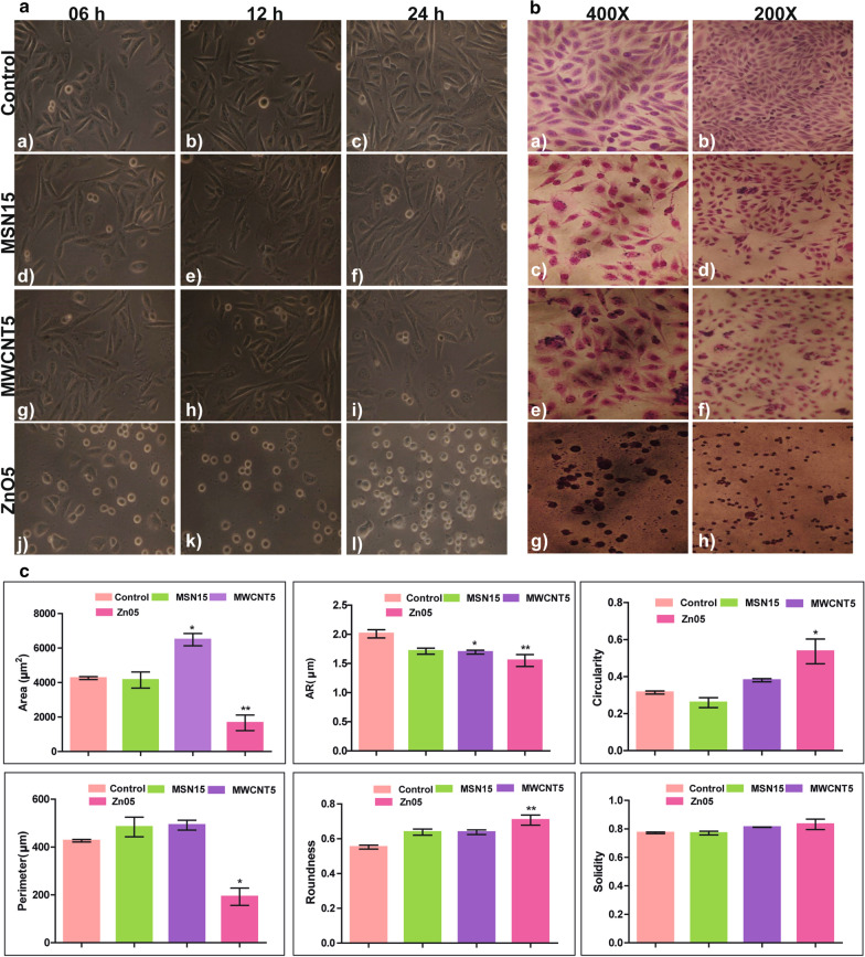
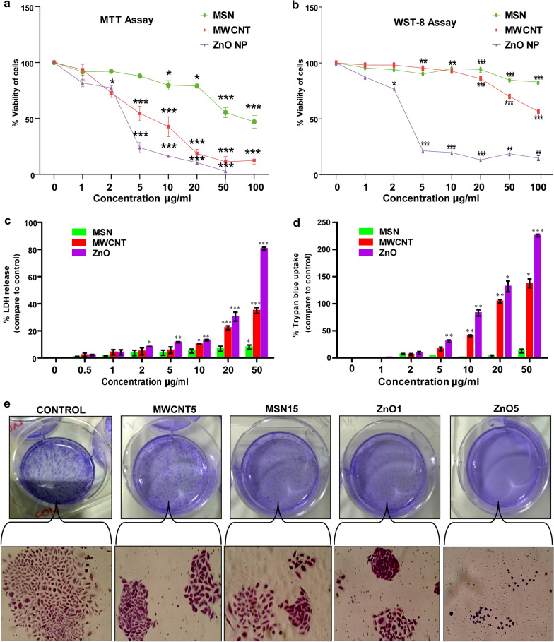
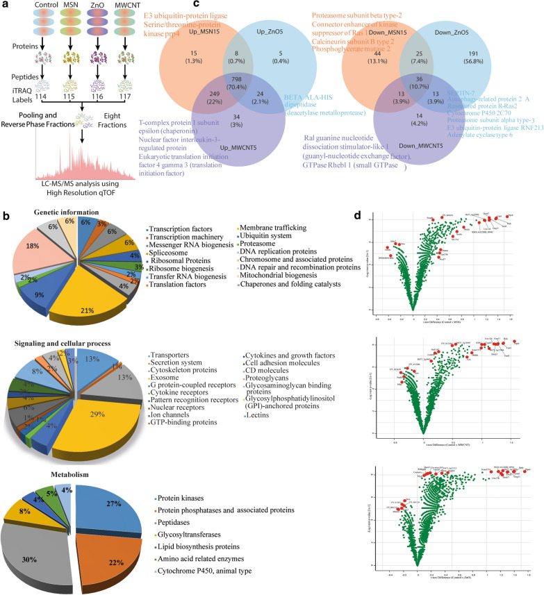
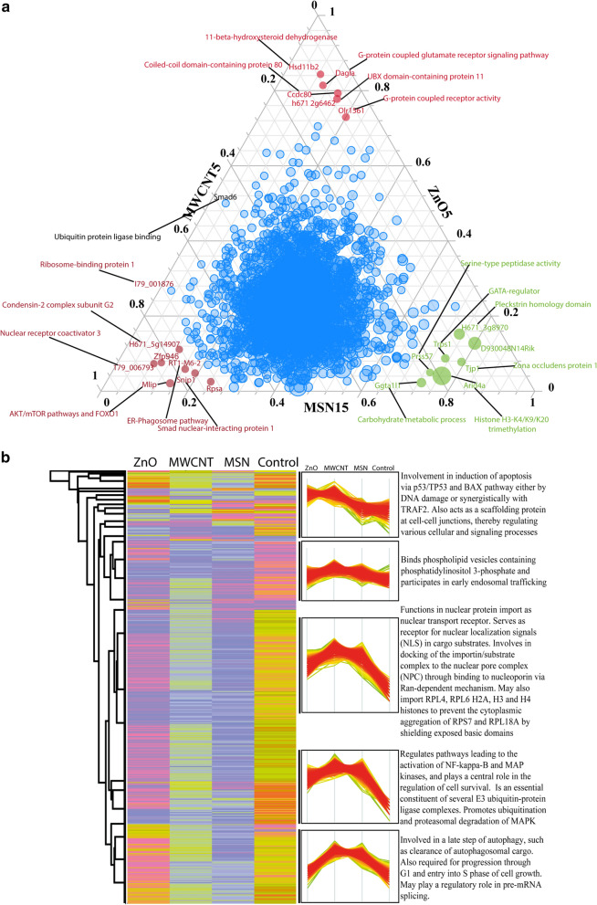
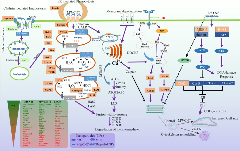
Through the high-throughput proteomic study, we observed that the effect of NPs is alone not restricted to cell viability but also on cell polarisation. In the case of MSN, no drastic changes were observed in cellular morphology, but it upregulated chaperons that might prevent protein aggregation. However, MWCNT showed elongated cell appearance with numerous cytoplasmic vacuoles, and induce lamellipodia formation through actin polymerisation. The cytoskeleton remodelling was accompanied by the increased expression of Dlc-1, cofilin and Rac1 proteins. While ZnO NPs resulted in the rounded cell morphology along with nuclear abnormalities. The proteome analysis revealed that UBXN11 control cell roundness and DOCK3 leads to actin stress fibre formation and finally, loss of cell adhesion. It enhances the expression of catastrophic DNA damage and apoptotic proteins, which was unrecoverable even after 72 h, as confirmed by the colony formation assay. All three NPs trigger over-expression of the endocytic pathway, ubiquitination, and proteasomal complex proteins. The data indicate that ZnO and MSN entered into the cells through clathrin-mediated pathways; whereas, MWCNT invades through ER-mediated phagocytosis.
Based on the incubation and concentration of NPs, our work provides evidence for the activation of Rac-Rho signalling pathway to alter cytoskeleton dynamics. Our results assist as a sensitive early molecular readout for nanosafety assessment.
目前尚未研究细胞对纳米颗粒(NPs)的机械线索和生化变化的反应。在这里,我们提供了对中国仓鼠卵巢(CHO-K1)细胞系的全面分析,以研究暴露于介孔硅纳米颗粒(MSN)、多壁碳纳米管(MWCNT)和氧化锌(ZnO) NPs 后细胞的行为。
通过高通量蛋白质组学研究,我们观察到 NPs 的影响不仅限于细胞活力,还影响细胞极化。在 MSN 的情况下,细胞形态没有明显变化,但它上调了伴侣蛋白,可能防止蛋白质聚集。然而,MWCNT 表现出伸长的细胞外观,并有许多细胞质空泡,并通过肌动蛋白聚合诱导片状伪足形成。细胞骨架重塑伴随着 Dlc-1、副肌球蛋白和 Rac1 蛋白的表达增加。而 ZnO NPs 导致细胞形态呈圆形,并伴有核异常。蛋白质组分析表明,UBXN11 控制细胞的圆形度,DOCK3 导致肌动蛋白应力纤维形成,最终导致细胞黏附丧失。它增强了灾难性 DNA 损伤和凋亡蛋白的表达,即使在 72 小时后也无法恢复,这一点通过集落形成实验得到了证实。这三种 NPs 都引发了内吞途径、泛素化和蛋白酶体复合物蛋白的过度表达。数据表明,ZnO 和 MSN 通过网格蛋白介导的途径进入细胞,而 MWCNT 通过内质网介导的吞噬作用进入细胞。
根据 NPs 的孵育和浓度,我们的工作为 Rac-Rho 信号通路的激活提供了证据,以改变细胞骨架动力学。我们的结果为纳米安全评估提供了敏感的早期分子读数。