Center for Environmental Implications of Nanotechnology, California NanoSystems Institute , University of California Los Angeles , 570 Westwood Plaza , Los Angeles , California 90095 , United States.
Division of NanoMedicine, Department of Medicine , University of California Los Angeles , 10833 Le Conte Ave. , Los Angeles , California 90095 , United States.
ACS Nano. 2018 Apr 24;12(4):3836-3852. doi: 10.1021/acsnano.8b01086. Epub 2018 Mar 19.
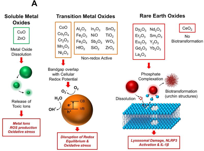
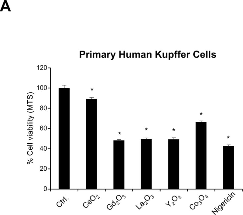
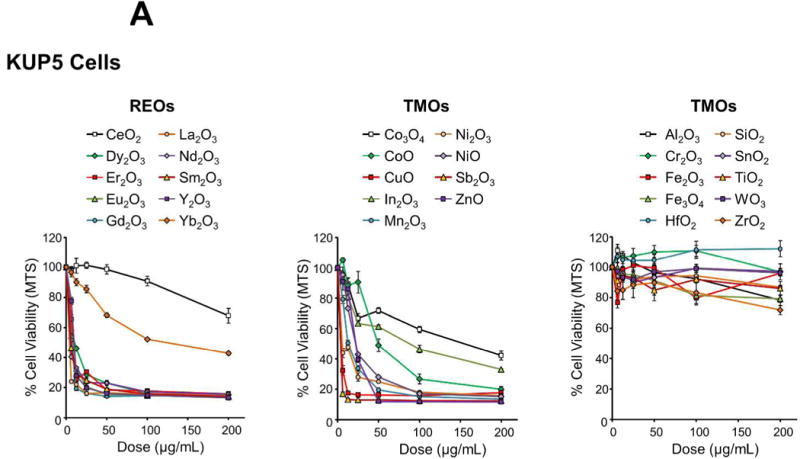
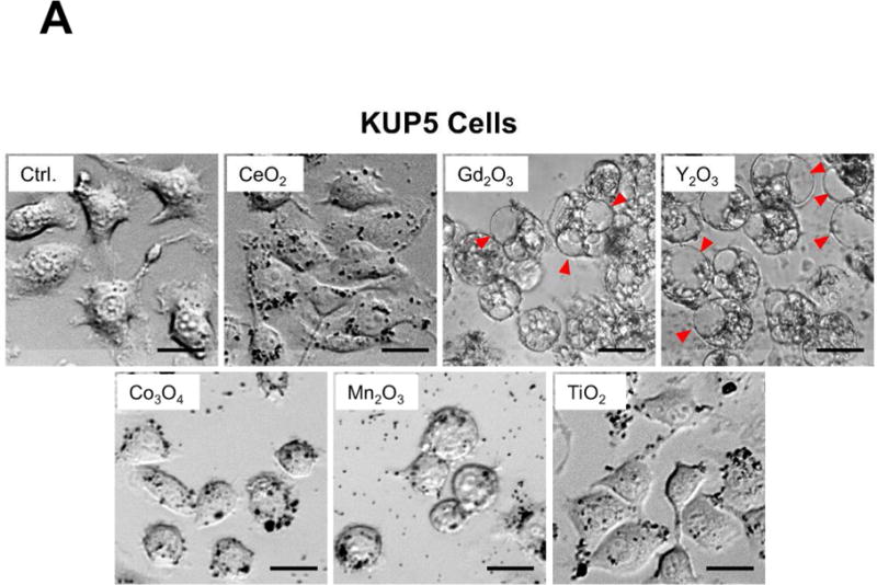
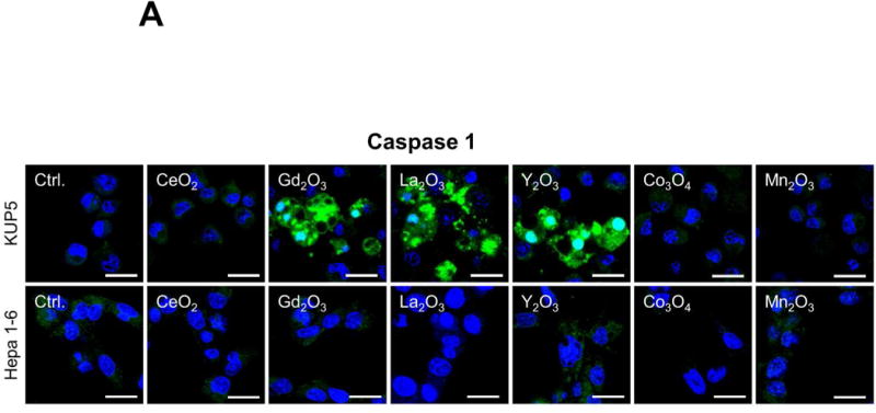
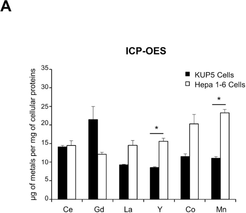
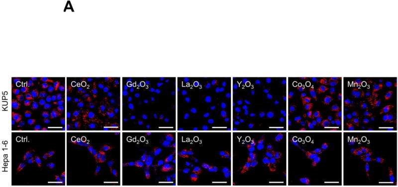
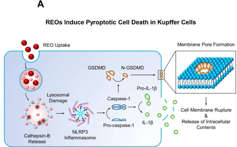
The liver and the mononuclear phagocyte system are a frequent target for engineered nanomaterials, either as a result of particle uptake and spread from primary exposure sites or systemic administration of therapeutic and imaging nanoparticles. In this study, we performed a comparative analysis of the toxicological impact of 29 metal oxide nanoparticles (NPs), some commonly used in consumer products, in transformed or primary Kupffer cells (KCs) and hepatocytes. We not only observed differences between KCs and hepatocytes, but also differences in the toxicological profiles of transition-metal oxides (TMOs, e. g., CoO) versus rare-earth oxide (REO) NPs ( e. g., GdO). While pro-oxidative TMOs induced the activation of caspases 3 and 7, resulting in apoptotic cell death in both cell types, REOs induced lysosomal damage, NLRP3 inflammasome activation, caspase 1 activation, and pyroptosis in KCs. Pyroptosis was accompanied by cell swelling, membrane blebbing, IL-1β release, and increased membrane permeability, which could be reversed by knockdown of the pore forming protein, gasdermin D. Though similar features were not seen in hepatocytes, the investigation of the cytotoxic effects of REO NPs could also be seen to affect macrophage cell lines such as J774A.1 and RAW 264.7 cells as well as bone marrow-derived macrophages. These phagocytic cell types also demonstrated features of pyroptosis and increased IL-1β production. Collectively, these findings demonstrate important mechanistic considerations that can be used for safety evaluation of metal oxides, including commercial products that are developed from these materials.
肝脏和单核吞噬细胞系统是工程纳米材料的常见靶标,这要么是由于颗粒摄取和从初始暴露部位扩散所致,要么是由于治疗和成像纳米颗粒的全身给药所致。在本研究中,我们对 29 种金属氧化物纳米颗粒(NPs)的毒理学影响进行了比较分析,这些纳米颗粒中的一些常用于消费产品,它们分别在转化或原代枯否细胞(KCs)和肝细胞中进行了研究。我们不仅观察到 KCs 和肝细胞之间存在差异,而且还观察到过渡金属氧化物(TMOs,例如 CoO)与稀土氧化物(REO)NPs(例如 GdO)的毒理学特征存在差异。虽然促氧化 TMOs 诱导了 caspase 3 和 7 的激活,导致两种细胞类型的凋亡性细胞死亡,但 REOs 诱导了溶酶体损伤、NLRP3 炎性体激活、caspase 1 激活和 KC 中的细胞焦亡。细胞焦亡伴随着细胞肿胀、细胞膜起泡、IL-1β释放和膜通透性增加,这些变化可以通过敲低孔形成蛋白 gasdermin D 来逆转。尽管在肝细胞中没有观察到类似的特征,但对 REO NPs 细胞毒性作用的研究也可以观察到对 J774A.1 和 RAW 264.7 细胞等巨噬细胞系以及骨髓来源的巨噬细胞的影响。这些吞噬细胞类型也表现出细胞焦亡和增加的 IL-1β 产生的特征。总之,这些发现表明了重要的机制考虑因素,这些因素可用于金属氧化物的安全性评估,包括由这些材料开发的商业产品。