Jiang Tao, Yan Yan, Zhou Kun, Su Chunxia, Ren Shengxiang, Li Nan, Hou Likun, Guo Xianchao, Zhu Wei, Zhang Henghui, Lin Jie, Zhang Jun, Zhou Caicun
Department of Medical Oncology, Shanghai Pulmonary Hospital & Thoracic Cancer Institute, Tongji University School of Medicine, 200433, Shanghai, China.
Department of Oncology, The First Affiliated Hospital of Zhengzhou University, 450052, Zhengzhou, China.
NPJ Precis Oncol. 2021 Feb 12;5(1):6. doi: 10.1038/s41698-021-00151-w.
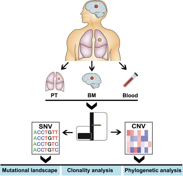
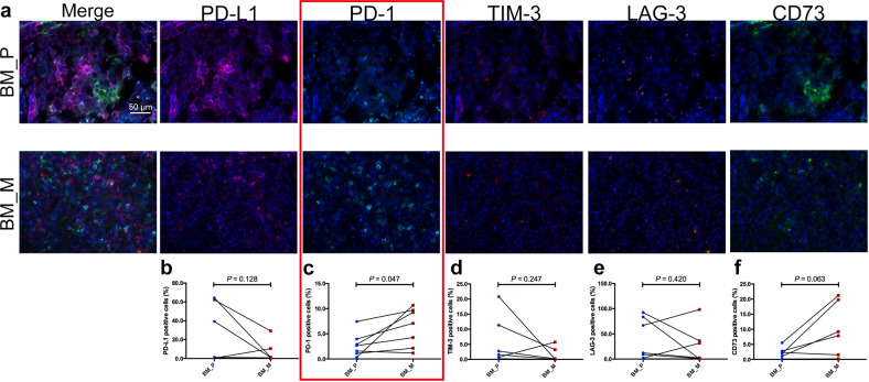
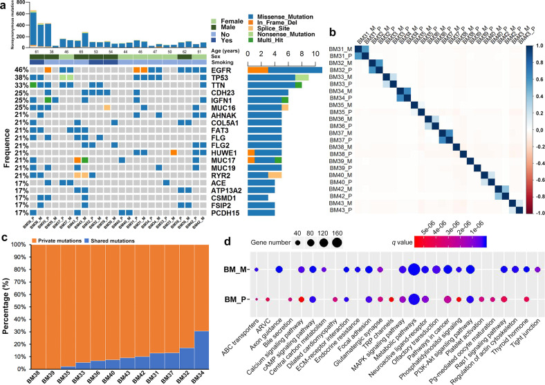
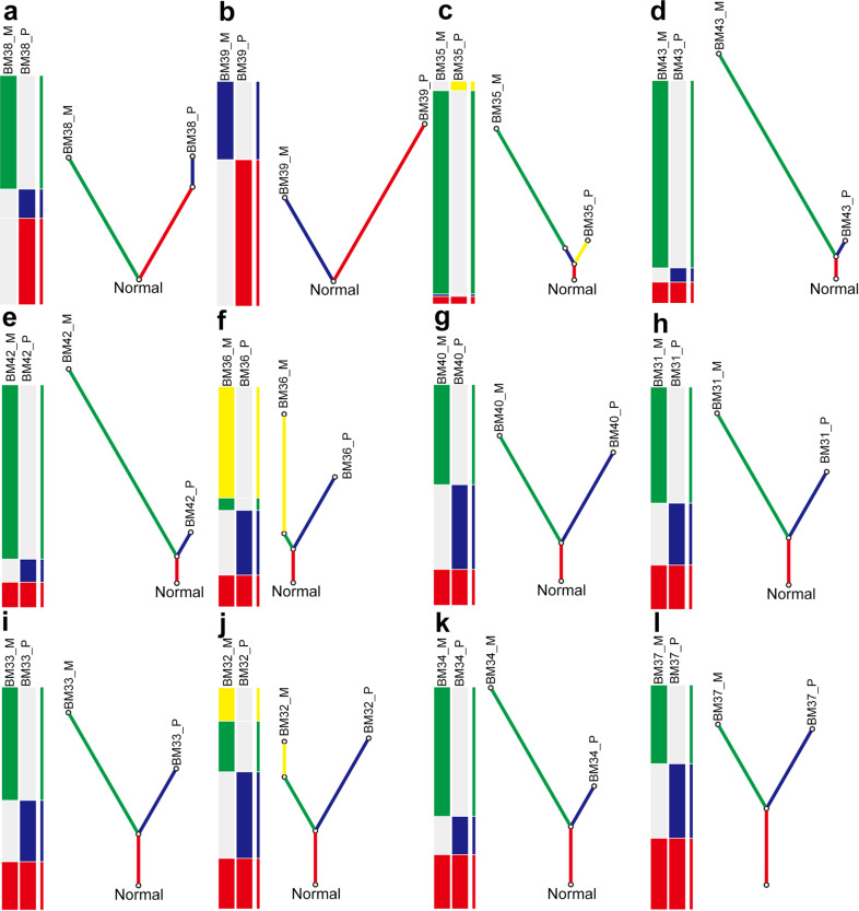
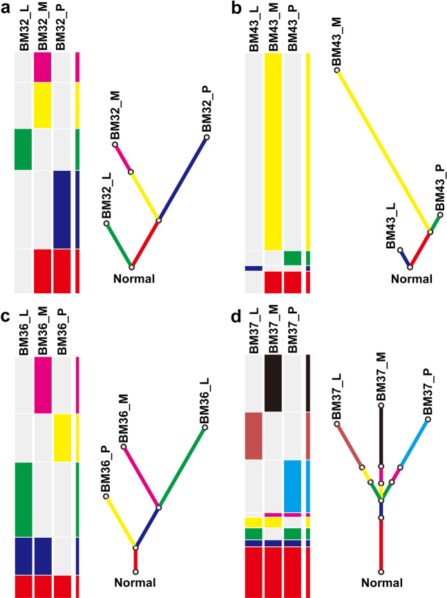
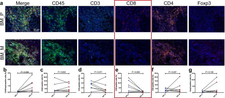
Characterizing the evolutionary trajectory and immune profiling of brain metastasis (BM) may provide insights in the development of novel therapeutic strategies. Here, we performed whole-exome sequencing and multiplex immunofluorescence (MIF) of 40 samples from 12 lung adenocarcinoma (LUAD) patients with BM and compared to their paired primary tumors. We observed significantly higher intertumor heterogeneity between paired primary tumors and BMs, with only a median of 8.3% of genetic mutations identified as shared. Phylogenetic analysis revealed that BM-competent clones genetically diverged from their primary tumors at relatively early stage, suggesting that the parallel progression model is dominant. In cases with synchronous lymph node metastasis (LNM), phylogenetic analysis suggested that BM is a later event than LNM. MIF analysis found that BMs exhibited significantly lower CD8 T cell infiltration (P = 0.048), and elevated CD4Foxp3 T cell infiltration (P = 0.036) and PD-1 expression (P = 0.047) in comparison to the matched primary tumors, indicating an immunosuppressive microenvironment in BMs. The current study revealed the discrepancy of mutational landscape as well as tumor immune microenvironment between BM and its primary tumor - such findings shall help us better understand the unique biological features of BM and develop innovative strategies accordingly for our patients with LUAD.
描绘脑转移(BM)的进化轨迹和免疫图谱可能为新型治疗策略的开发提供见解。在此,我们对12例患有脑转移的肺腺癌(LUAD)患者的40个样本进行了全外显子组测序和多重免疫荧光(MIF)分析,并与它们配对的原发性肿瘤进行了比较。我们观察到配对的原发性肿瘤和脑转移瘤之间的肿瘤间异质性显著更高,只有中位数8.3%的基因突变被确定为共享。系统发育分析表明,具有脑转移能力的克隆在相对早期就与其原发性肿瘤发生了基因分歧,这表明平行进展模型占主导地位。在伴有同步淋巴结转移(LNM)的病例中,系统发育分析表明脑转移是比淋巴结转移更晚发生的事件。MIF分析发现,与匹配的原发性肿瘤相比,脑转移瘤表现出显著更低的CD8 T细胞浸润(P = 0.048),以及升高的CD4Foxp3 T细胞浸润(P = 0.036)和PD-1表达(P = 0.047),这表明脑转移瘤中存在免疫抑制微环境。当前研究揭示了脑转移与其原发性肿瘤之间在突变格局以及肿瘤免疫微环境方面的差异——这些发现将有助于我们更好地理解脑转移的独特生物学特征,并相应地为我们的LUAD患者制定创新策略。