Department of Pharmacology, University of the Basque Country UPV/EHU, Leioa, Spain.
Glial Cell Biology Lab, Achucarro Basque Center for Neuroscience, Leioa, Spain.
Front Immunol. 2021 Jan 29;11:620602. doi: 10.3389/fimmu.2020.620602. eCollection 2020.
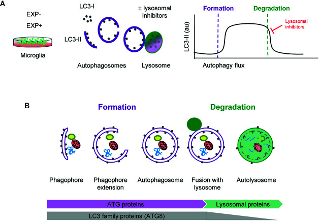
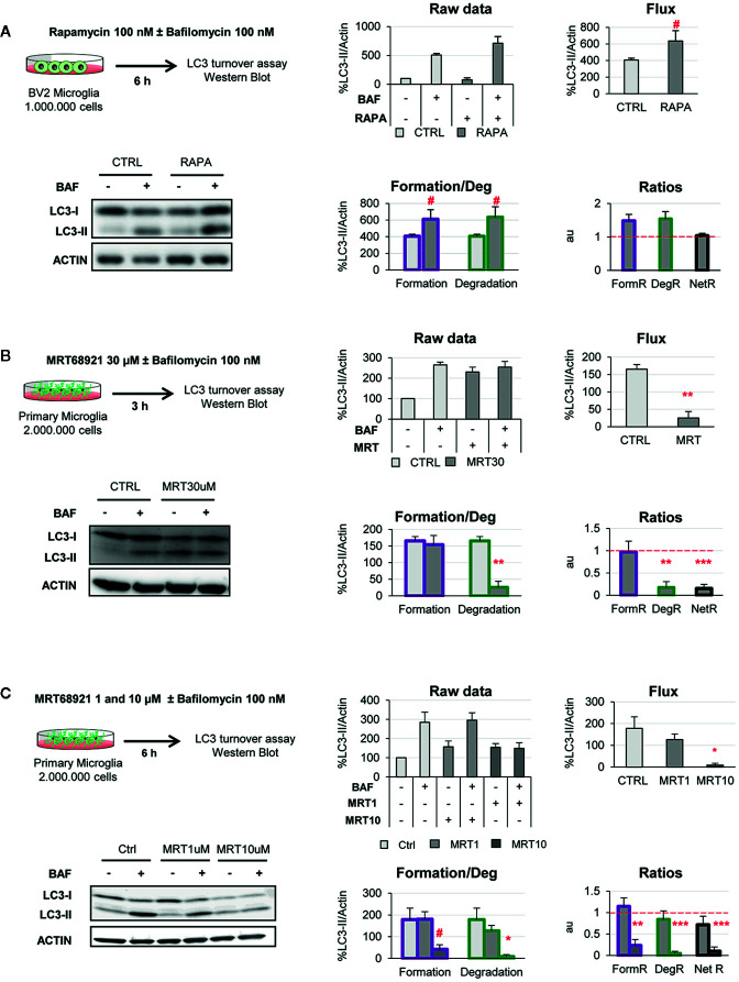
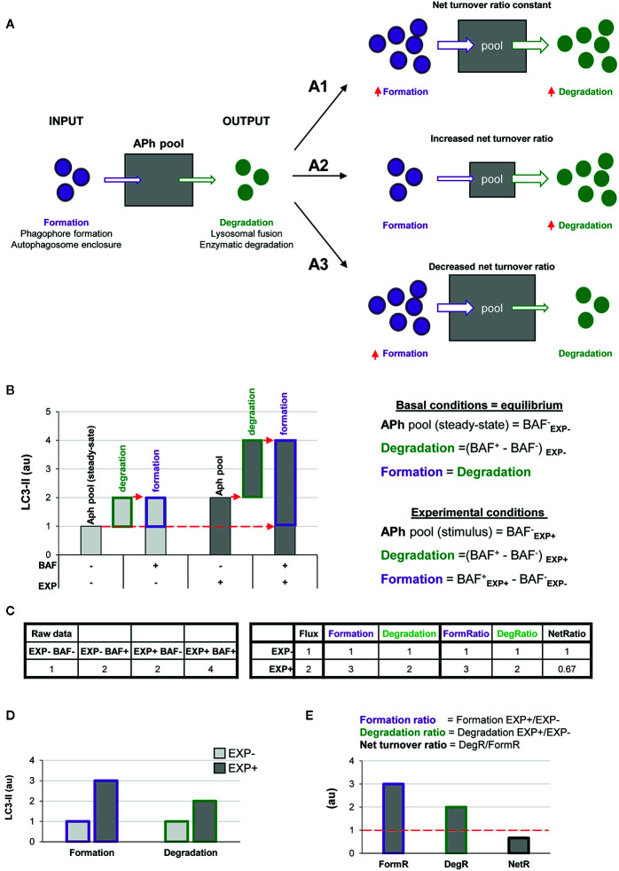
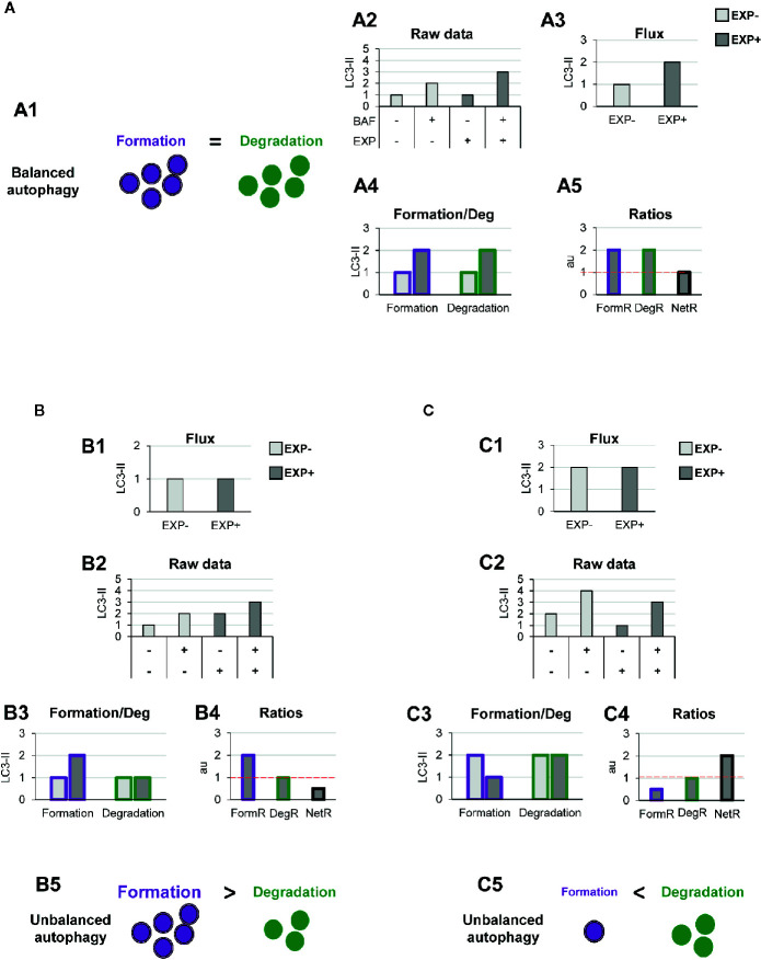
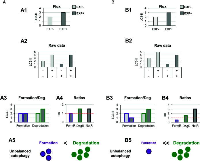
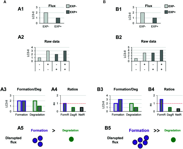
Autophagy is a complex process that encompasses the enclosure of cytoplasmic debris or dysfunctional organelles in membranous vesicles, the autophagosomes, for their elimination in the lysosomes. Autophagy is increasingly recognized as a critical process in macrophages, including microglia, as it finely regulates innate immune functions such as inflammation. A gold-standard method to assess its induction is the analysis of the autophagic flux using as a surrogate the expression of the microtubule-associated light chain protein 3 conjugated to phosphatidylethanolamine (LC3-II) by Western blot, in the presence of lysosomal inhibitors. Therefore, the current definition of autophagy flux actually puts the focus on the degradation stage of autophagy. In contrast, the most important autophagy controlling genes that have been identified in the last few years in fact target early stages of autophagosome formation. From a biological standpoint is therefore conceivable that autophagosome formation and degradation are independently regulated and we argue that both stages need to be systematically analyzed. Here, we propose a simple two-step model to understand changes in autophagosome formation and degradation using data from conventional LC3-II Western blot, and test it using two models of autophagy modulation in cultured microglia: rapamycin and the ULK1/2 inhibitor, MRT68921. Our two-step model will help to unravel the effect of genetic, pharmacological, and environmental manipulations on both formation and degradation of autophagosomes, contributing to dissect out the role of autophagy in physiology and pathology in microglia as well as other cell types.
自噬是一个复杂的过程,它包括将细胞质碎片或功能失调的细胞器包裹在膜泡中,即自噬体中,以便在溶酶体中进行消除。自噬在巨噬细胞中,包括小神经胶质细胞中,被越来越多地认为是一个关键过程,因为它精细地调节了先天免疫功能,如炎症。评估其诱导的金标准方法是使用Western blot 分析微管相关轻链蛋白 3 与磷脂酰乙醇胺(LC3-II)的共轭物的自噬通量,同时存在溶酶体抑制剂。因此,目前自噬通量的定义实际上侧重于自噬的降解阶段。相比之下,近年来鉴定的最重要的自噬控制基因实际上针对自噬体形成的早期阶段。因此,从生物学角度来看,可以想象自噬体的形成和降解是独立调节的,我们认为这两个阶段都需要系统地分析。在这里,我们提出了一个简单的两步模型,使用常规 LC3-II Western blot 中的数据来理解自噬体形成和降解的变化,并使用培养的小神经胶质细胞中两种自噬调节模型(雷帕霉素和 ULK1/2 抑制剂 MRT68921)对其进行测试。我们的两步模型将有助于揭示遗传、药理学和环境操作对自噬体形成和降解的影响,有助于阐明自噬在小神经胶质细胞以及其他细胞类型中的生理和病理中的作用。