Pierre Alexandre, Favory Raphael, Brassart Benoit, Bourel Claire, Romien Raphael, Normandin Sylvain, Dubech Arthur, Vincent Claire, Lemaire Jeremy, Grolaux Gaelle, Not Ophelie, Wallet Frederic, Daussin Frederic, Boulanger Eric, Durand Arthur, Frimat Marie, Ghinet Alina, Howsam Michael, Laine William, Marchetti Philippe, Kluza Jerome, Chatelain Estelle, Vandel Jimmy, Barois Nicolas, Montel Valerie, Bastide Bruno, Preau Sebastien, Lancel Steve
Univ. Lille, Inserm, CHU Lille, Institut Pasteur de Lille, U1167-RID-AGE-Facteurs de Risque et Déterminants Moléculaires des Maladies Liées au Vieillissement, Lille, France.
Division of Intensive Care, Hôpital Roger Salengro, CHU de Lille, Lille, France.
J Cachexia Sarcopenia Muscle. 2025 Aug;16(4):e70041. doi: 10.1002/jcsm.70041.
Sepsis survivors often experience sustained muscle weakness, leading to physical disability, with no pharmacological treatments available. Despite these well-documented long-term clinical consequences, research exploring the cellular and molecular mechanisms is sorely lacking.
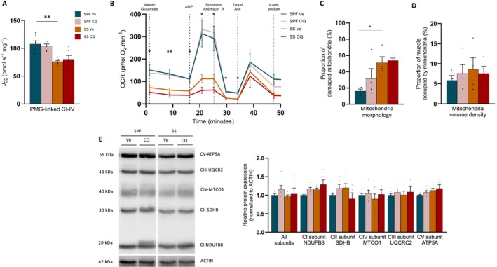
Bioinformatic analysis was performed in the vastus lateralis transcriptome of human ICU survivors 7 days after ICU discharge (D7), 6 months (M6) and age- and sex-matched controls. Enrichment analysis using Gene Ontology (GO) terms and Mitocarta3.0 was performed at D7 and M6 on differentially expressed genes (DEGs) and modules identified by weighted gene co-expression network analysis (WGCNA). Using a murine model of resuscitated sepsis induced by caecal slurry injection, pathways identified by the bioinformatics analysis were explored in 18- to 24-week-old sepsis-surviving (SS) mice at Day 10. Autophagy flux was investigated both in vivo and in vitro with chloroquine, a lysosomal inhibitor and urolithin A (UA), an autophagy inducer. Systemic metabolism was evaluated with indirect calorimetry, muscle phenotype with in situ and ex vivo contractility, muscle mass, myofibre cross-sectional area and typing and mitochondrial population with transmission electron microscopy (TEM), as well as mitochondrial function with high-resolution respirometry. Autophagic vacuole (AV) level was monitored using LC3B-II and P62 protein expression and TEM.
Pathways related to 'mitochondrion' were the only ones whose deregulation persisted between D7 and M6 (p < 0.05) and characterized WGCNA modules correlated with muscle mass, strength and physical function. Shared mitochondrial DEGs between D7 and M6 encoded matrix mitochondrial proteins related to 'metabolism' and 'mitochondrial dynamics'. SS mice exhibited reduced complex I-driven oxygen consumption (CI-J) (-45%), increased S-nitrosylation of complex I, damaged (+35%) and oxidized (+51%) mitochondria and AV accumulation (5 vs. 50 AVs/mm) compared with sham pair-fed mice (p < 0.05) despite no differences in mitochondrial size or number. Autophagy flux was reduced in SS mice due to decreased AV degradation ratio (p < 0.05). UA restored a balanced autophagy flux (turnover ratio 0.96 vs. -0.17) by increasing AVs formation and degradation ratio (p < 0.05). UA also improved CI-J (81 vs. 106 pmol/s/mg), tetanic force (215 vs. 244 mN/mm) and hindlimb muscle weight in SS mice (p < 0.05).
Mitochondrial and autophagy disruption contributes to long-term muscle dysfunction in human and mouse sepsis survivors. We demonstrate for the first time that sepsis induces an autophagy flux blockade. Urolithin A prevents mitochondrial and muscle impairments both in vivo and in vitro by improving autophagy flux.
脓毒症幸存者常经历持续性肌肉无力,导致身体残疾,且尚无可用的药物治疗方法。尽管这些长期临床后果已有充分记录,但探索其细胞和分子机制的研究却严重不足。
对人类重症监护病房(ICU)幸存者在出院7天(D7)、6个月(M6)时的股外侧肌转录组以及年龄和性别匹配的对照组进行了生物信息学分析。使用基因本体论(GO)术语和Mitocarta3.0对D7和M6时通过加权基因共表达网络分析(WGCNA)鉴定的差异表达基因(DEG)和模块进行富集分析。使用盲肠灌注诱导的复苏型脓毒症小鼠模型,在第10天对18至24周龄的脓毒症存活(SS)小鼠中通过生物信息学分析确定的通路进行研究。使用溶酶体抑制剂氯喹和自噬诱导剂尿石素A(UA)在体内和体外研究自噬通量。通过间接测热法评估全身代谢,通过原位和离体收缩性、肌肉质量、肌纤维横截面积和类型以及透射电子显微镜(TEM)观察线粒体数量来评估肌肉表型,并通过高分辨率呼吸测定法评估线粒体功能。使用LC3B-II和P62蛋白表达以及TEM监测自噬泡(AV)水平。
与“线粒体”相关的通路是唯一在D7和M6之间持续失调(p < 0.05)且表征与肌肉质量、力量和身体功能相关的WGCNA模块的通路。D7和M6之间共享的线粒体DEG编码与“代谢”和“线粒体动力学”相关的线粒体基质蛋白。与假手术配对喂养的小鼠相比,SS小鼠表现出复合物I驱动的氧消耗(CI-J)降低(-45%),复合物I的S-亚硝基化增加,线粒体受损(+35%)和氧化(+51%)以及AV积累(5个对50个AVs/mm)(p < 0.05),尽管线粒体大小或数量没有差异。由于AV降解率降低,SS小鼠的自噬通量减少(p < 0.05)。UA通过增加AV形成和降解率恢复了平衡的自噬通量(周转率0.96对-0.17)(p < 0.05)。UA还改善了SS小鼠的CI-J(81对106 pmol/s/mg)、强直力(215对244 mN/mm)和后肢肌肉重量(p < 0.05)。
线粒体和自噬破坏导致人类和小鼠脓毒症幸存者出现长期肌肉功能障碍。我们首次证明脓毒症诱导自噬通量阻断。尿石素A通过改善自噬通量在体内和体外预防线粒体和肌肉损伤。