Department of Cardiology, Barts Heart Centre, Barts Health NHS Trust, West Smithfield, London, EC1A 7BE, UK.
Centre for Cardiovascular Medicine and Devices, William Harvey Research Institute, Queen Mary University of London, London, UK.
Int J Cardiovasc Imaging. 2021 Jun;37(6):1825-1837. doi: 10.1007/s10554-021-02162-x. Epub 2021 Feb 15.
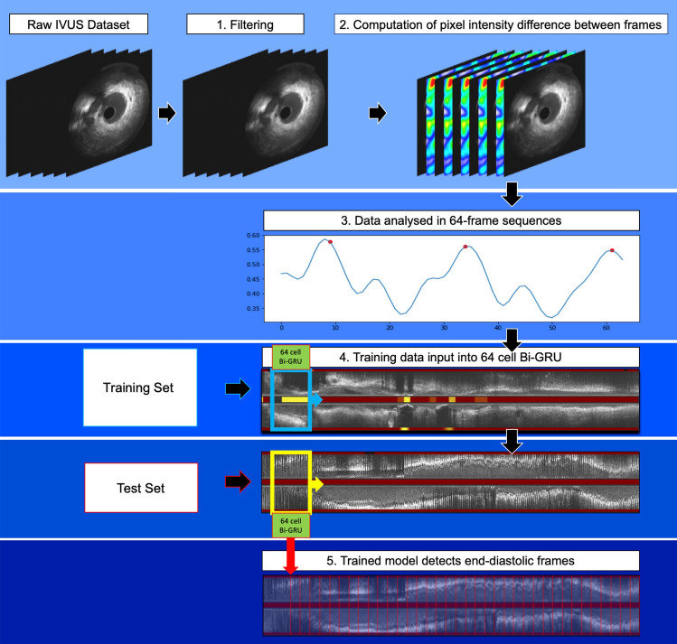
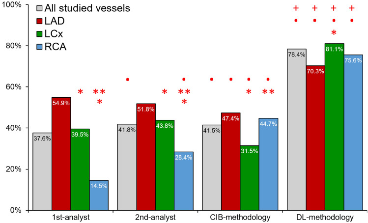
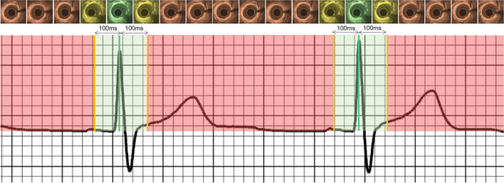
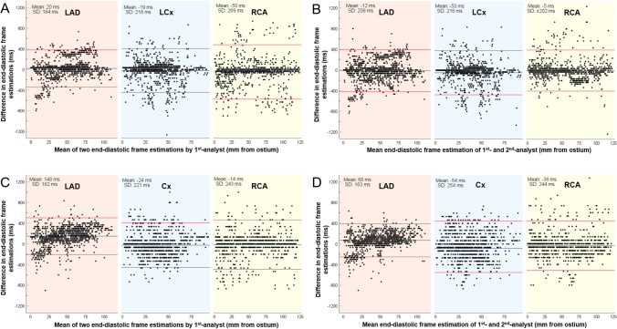
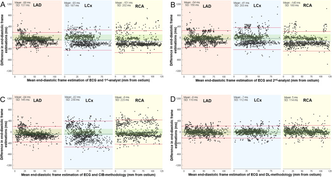
Coronary luminal dimensions change during the cardiac cycle. However, contemporary volumetric intravascular ultrasound (IVUS) analysis is performed in non-gated images as existing methods to acquire gated or to retrospectively gate IVUS images have failed to dominate in research. We developed a novel deep learning (DL)-methodology for end-diastolic frame detection in IVUS and compared its efficacy against expert analysts and a previously established methodology using electrocardiographic (ECG)-estimations as reference standard. Near-infrared spectroscopy-IVUS (NIRS-IVUS) data were prospectively acquired from 20 coronary arteries and co-registered with the concurrent ECG-signal to identify end-diastolic frames. A DL-methodology which takes advantage of changes in intensity of corresponding pixels in consecutive NIRS-IVUS frames and consists of a network model designed in a bidirectional gated-recurrent-unit (Bi-GRU) structure was trained to detect end-diastolic frames. The efficacy of the DL-methodology in identifying end-diastolic frames was compared with two expert analysts and a conventional image-based (CIB)-methodology that relies on detecting vessel movement to estimate phases of the cardiac cycle. A window of ± 100 ms from the ECG estimations was used to define accurate end-diastolic frames detection. The ECG-signal identified 3,167 end-diastolic frames. The mean difference between DL and ECG estimations was 3 ± 112 ms while the mean differences between the 1st-analyst and ECG, 2nd-analyst and ECG and CIB-methodology and ECG were 86 ± 192 ms, 78 ± 183 ms and 59 ± 207 ms, respectively. The DL-methodology was able to accurately detect 80.4%, while the two analysts and the CIB-methodology detected 39.0%, 43.4% and 42.8% of end-diastolic frames, respectively (P < 0.05). The DL-methodology can identify NIRS-IVUS end-diastolic frames accurately and should be preferred over expert analysts and CIB-methodologies, which have limited efficacy.
冠状动脉管腔在心动周期中会发生变化。然而,目前的容积血管内超声(IVUS)分析是在非门控图像中进行的,因为现有的获取门控或回顾性门控 IVUS 图像的方法在研究中都未能占据主导地位。我们开发了一种新的深度学习(DL)方法来检测 IVUS 中的舒张末期帧,并将其与专家分析者和以前使用心电图(ECG)估计值作为参考标准的方法进行了比较。近红外光谱-IVUS(NIRS-IVUS)数据从 20 条冠状动脉前瞻性采集,并与同步的 ECG 信号进行配准,以识别舒张末期帧。一种利用连续 NIRS-IVUS 帧中相应像素强度变化的 DL 方法,由一个双向门控循环单元(Bi-GRU)结构设计的网络模型组成,用于检测舒张末期帧。该 DL 方法识别舒张末期帧的效果与两名专家分析者和一种基于传统图像(CIB)的方法进行了比较,该方法依赖于检测血管运动来估计心动周期的相位。使用 ECG 估计值的±100ms 窗口来定义准确的舒张末期帧检测。ECG 信号识别了 3167 个舒张末期帧。DL 和 ECG 估计之间的平均差异为 3±112ms,而第一分析者和 ECG、第二分析者和 ECG 以及 CIB 方法和 ECG 之间的平均差异分别为 86±192ms、78±183ms 和 59±207ms。DL 方法能够准确地检测到 80.4%,而两名分析者和 CIB 方法分别检测到 39.0%、43.4%和 42.8%的舒张末期帧(P<0.05)。DL 方法能够准确地识别 NIRS-IVUS 的舒张末期帧,应优先于专家分析者和 CIB 方法,后者的效果有限。