Department of Pathophysiology, College of Basic Medical Science, China Medical University, No.77, Puhe Road, Shenyang North New Area, Shenyang, 110122, Liaoning Province, China.
Department of Pathophysiology, Zhangjiakou University, No.P19, Pingmen Street, Qiaoxi District, Zhangjiakou, 075000, Hebei Province, China.
Stem Cell Res Ther. 2021 Feb 17;12(1):141. doi: 10.1186/s13287-021-02192-1.
As a large capillary network, the human placenta plays an important role throughout pregnancy. Placental vascular development is complex and delicate and involves many types of placental cells, such as trophoblasts, and mesenchymal stem cells. There has been no systematic, comparative study on the roles of these two groups of placental cells and the whole placental tissue in the placental angiogenesis. In this study, primary cytotrophoblasts (CTBs) from early pregnancy and primary human placenta-derived mesenchymal stem cells (hPDMSCs) from different stages of pregnancy were selected as the cell research objects, and full-term placental tissue was selected as the tissue research object to detect the effects of their conditioned medium (CM) on human umbilical vein endothelial cell (HUVEC) angiogenesis.
We successfully isolated primary hPDMSCs and CTBs, collected CM from these placental cells and sub-cultured placental tissue, and then evaluated the effects of the CM on a series of angiogenic processes in HUVECs in vitro. Furthermore, we measured the levels of angiogenic factors in the CM of placental cells or tissue by an angiogenesis antibody array.
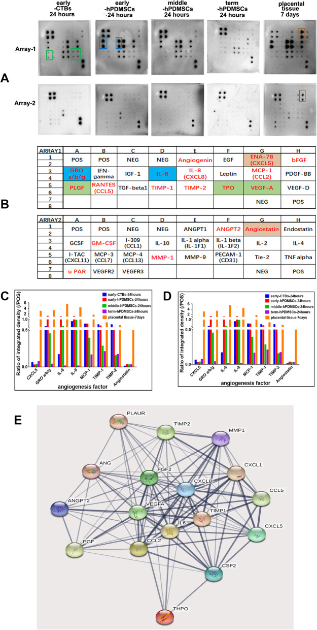
The results showed that not only placental cells but also sub-cultured placental tissue, to some extent, promoted HUVEC angiogenesis in vitro by promoting proliferation, adhesion, migration, invasion, and tube formation. We also found that primary placental cells in early pregnancy, whether CTBs or hPDMSCs, played more significant roles than those in full-term pregnancy. Placental cell-derived CM collected at 24 h or 48 h had the best effect, and sub-cultured placental tissue-derived CM collected at 7 days had the best effect among all the different time points. The semiquantitative angiogenesis antibody array showed that 18 of the 43 angiogenic factors had obvious spots in placental cell-derived CM or sub-cultured placental tissue-derived CM, and the levels of 5 factors (including CXCL-5, GRO, IL-6, IL-8, and MCP-1) were the highest in sub-cultured placental tissue-derived CM.
CM obtained from placental cells (primary CTBs or hPDMSCs) or sub-cultured placental tissue contained proangiogenic factors and promoted HUVEC angiogenesis in vitro. Therefore, our research is helpful to better understand placental angiogenesis regulation and provides theoretical support for the clinical application of placental components, especially sub-cultured placental tissue-derived CM, in vascular tissue engineering and clinical treatments.
作为一个大型毛细血管网络,胎盘在整个孕期中起着重要作用。胎盘血管发育复杂而微妙,涉及多种胎盘细胞,如滋养细胞和间充质干细胞。目前还没有系统的、比较性的研究来评估这两组胎盘细胞和整个胎盘组织在胎盘血管生成中的作用。在这项研究中,选择早孕的原代滋养细胞(CTBs)和不同孕期的原代人胎盘来源间充质干细胞(hPDMSCs)作为细胞研究对象,选择足月胎盘组织作为组织研究对象,检测其条件培养基(CM)对人脐静脉内皮细胞(HUVEC)血管生成的影响。
我们成功分离了原代 hPDMSCs 和 CTBs,收集了这些胎盘细胞的 CM 并进行了胎盘组织的传代培养,然后评估了 CM 对 HUVEC 体外一系列血管生成过程的影响。此外,我们通过血管生成抗体阵列测量了胎盘细胞或组织 CM 中的血管生成因子水平。
结果表明,不仅胎盘细胞,而且传代培养的胎盘组织在一定程度上通过促进增殖、黏附、迁移、侵袭和管形成促进了体外 HUVEC 血管生成。我们还发现,无论是 CTBs 还是 hPDMSCs,早孕的原代胎盘细胞比足月妊娠的胎盘细胞发挥的作用更为显著。在所有不同的时间点中,24 小时或 48 小时收集的胎盘细胞衍生的 CM 效果最佳,而在所有不同的时间点中,7 天收集的传代培养胎盘组织衍生的 CM 效果最佳。半定量血管生成抗体阵列显示,43 种血管生成因子中有 18 种在胎盘细胞衍生的 CM 或传代培养的胎盘组织衍生的 CM 中有明显的斑点,其中 5 种因子(包括 CXCL-5、GRO、IL-6、IL-8 和 MCP-1)在传代培养的胎盘组织衍生的 CM 中的水平最高。
胎盘细胞(原代 CTBs 或 hPDMSCs)或传代培养的胎盘组织获得的 CM 含有促血管生成因子,并在体外促进 HUVEC 血管生成。因此,我们的研究有助于更好地理解胎盘血管生成的调节,并为胎盘成分(特别是传代培养的胎盘组织衍生的 CM)在血管组织工程和临床治疗中的临床应用提供理论支持。