Ewelina Kazimierczyk, Eljaszewicz Andrzej, Kazimierczyk Remigiusz, Tynecka Marlena, Zembko Paula, Tarasiuk Ewa, Kaminski Karol, Sobkowicz Bozena, Moniuszko Marcin, Tycinska Agnieszka
Department of Cardiology, Medical University of Bialystok, Bialystok, Poland.
Department of Regenerative Medicine and Immune Regulation, Medical University of Bialystok, Bialystok, Poland.
Postepy Kardiol Interwencyjnej. 2020 Sep;16(3):287-293. doi: 10.5114/aic.2020.99263. Epub 2020 Oct 2.
In the course of acute myocardial infarction (AMI) cardiomyocyte injury, activation and destruction of endothelial cells together with inflammation lead to miRNA expression alterations.
To assess levels of circulating cardiac-specific (miR-1) and endothelial-specific (miR-126) miRNAs in the acute phase of AMI and after a follow-up period.
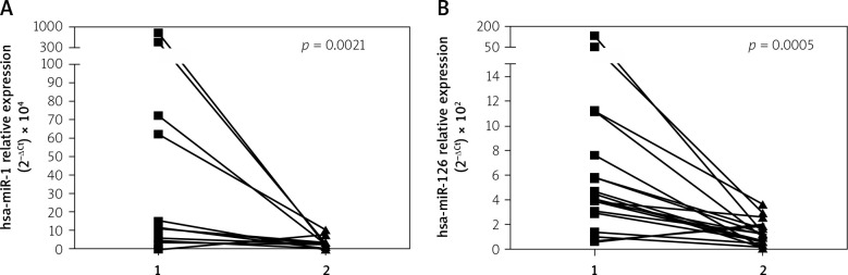
Seventeen AMI patients (mean age: 64.24 ±13.83 years, mean left ventricle ejection fraction (LVEF): 42.6 ±9.65%), treated with primary percutaneous coronary intervention within the first 12 h, had plasma miRNAs isolated (quantitative real-time PCR, Exiqon) on admission and after 19.2 ±5.9 weeks. Measurements were also performed in a control group of healthy volunteers matched for age and sex.
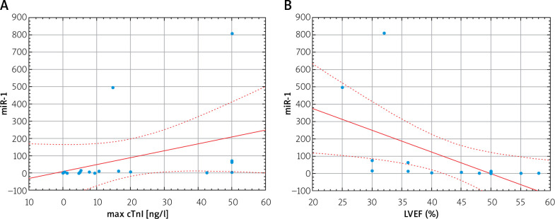
Concentrations of both miRNAs were significantly higher in AMI patients as compared to healthy controls: miR-1: 5.93 (3.15-14.92) vs. 1.46 (0.06-2.96), = 0.04; miR-126: 4.5 (3.11-7.64) vs. 0.54 (0.36-0.99), = 0.00003, respectively. Levels of both miRNAs significantly decreased after the follow-up period: miR-1: 5.93 (3.15-14.92) vs. 1.34 (0.04-2.34), = 0.002; miR-126: 4.5 (3.11-7.64) vs. 1.18 (0.49-1.68), = 0.0005). Moreover, miR-1 correlated positively with maximal troponin I concentration ( = 0.59, = 0.02) and negatively with LVEF ( = -0.76, = 0.0004).
In our study, miR-1 emerged as a marker of cardiomyocyte injury and loss of myocardial contractility, whereas dynamics of miR-126 concentration may reflect endothelial activation and damage in the most extreme stage of atherosclerosis, followed by angiogenesis in ischemic myocardium. However, to fully elucidate the role of miR-1 and miR-126 as biomarkers of AMI and future therapeutic targets, further research is required.
在急性心肌梗死(AMI)过程中,心肌细胞损伤、内皮细胞激活与破坏以及炎症反应共同导致微小RNA(miRNA)表达改变。
评估急性心肌梗死急性期及随访期循环中心脏特异性(miR-1)和内皮细胞特异性(miR-126)miRNA的水平。
17例急性心肌梗死患者(平均年龄:64.24±13.83岁,平均左心室射血分数(LVEF):42.6±9.65%),在发病后12小时内接受了直接经皮冠状动脉介入治疗,入院时及19.2±5.9周后采集血浆分离miRNA(定量实时聚合酶链反应,Exiqon公司产品)。同时在年龄和性别匹配的健康志愿者对照组中进行检测。
与健康对照组相比急性心肌梗死患者中两种miRNA的浓度均显著升高:miR-1:5.93(3.15 - 14.92)对1.46(0.06 - 2.96),P = 0.04;miR-126:4.5(3.11 - 7.64)对0.54(0.36 - 0.99),P = 0.00003。随访期后两种miRNA水平均显著下降:miR-1:5.93(3.15 - 14.92)对1.34(0.04 - 2.34),P = 0.002;miR-126:4.5(3.11 - 7.64)对1.18(0.49 - 1.68),P = 0.0005。此外,miR-1与肌钙蛋白I最大浓度呈正相关(r = 0.59,P = 0.02),与左心室射血分数呈负相关(r = -0.76,P = 0.0004)。
在我们的研究中,miR-1成为心肌细胞损伤和心肌收缩力丧失的标志物,而miR-126浓度的动态变化可能反映动脉粥样硬化最严重阶段的内皮细胞激活和损伤,随后是缺血心肌的血管生成。然而,要全面阐明miR-1和miR-126作为急性心肌梗死生物标志物及未来治疗靶点的作用,还需要进一步研究。