Amin T N, Wong M, Foo X, Pointer S-L, Goodhart V, Jurkovic D
Institute for Women's Health, University College London Hospitals NHS Foundation Trust, 250 Euston Road, London, NW1 2PG, UK.
Ultrasound J. 2021 Feb 18;13(1):7. doi: 10.1186/s13089-021-00212-y.
Transvaginal ultrasound (TVS) is a sensitive tool for detecting various conditions that contribute to pelvic pain. TVS can be also used to assess blood flow and measure the size of pelvic veins. Pelvic venous congestion (PVC) is characterised by enlargement of the pelvic veins and has been recognised as a cause of chronic pelvic pain. The reference ranges for uterine venous diameter in women with normal pelvic organs have been established, but there is no information regarding the potential effect of pelvic pathology on the uterine venous diameters. The aim of this study was to examine the size of uterine venous plexus in women with evidence of pelvic abnormalities on TVS and to determine whether the reference ranges need to be adjusted in the presence of pelvic pathology. A prospective, observational study was conducted in our gynaecological outpatient clinic. Morphological characteristics of all pelvic abnormalities detected on TVS and their sizes were recorded. The uterine veins were identified and their diameters were measured in all cases. The primary outcome measure was the uterine venous diameter. Regression analyses were performed to determine factors affecting the uterine venous size in women with pelvic pathology.
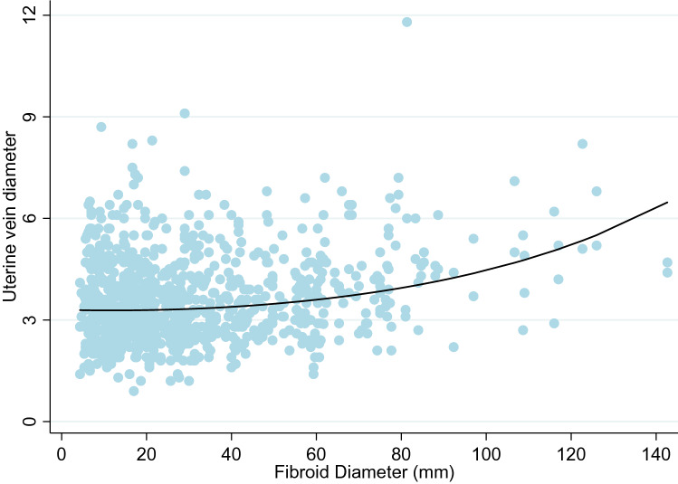
A total of 1500 women were included into the study, 1014 (67%) of whom were diagnosed with pelvic abnormalities. Women with pelvic pathology had significantly larger uterine venous diameters than women with normal pelvic organs (p < 0.01). Multivariable analysis showed that pre-menopausal status, high parity, presence of fibroids (p < 0.001) and Black ethnicity were all associated with significantly larger uterine vein diameters. Based on these findings modified reference ranges for uterine venous diameters have been designed which could be used for the diagnosis of PVC in women with uterine fibroids.
Our findings show that of all pelvic pathology detected on TVS, only fibroids are significantly associated with uterine venous enlargement. Factors known to be associated with enlarged veins in women with normal pelvic organs, namely parity and menopausal status, also apply in patients with pelvic pathology. Future studies of uterine venous circulation should take into account the presence and size of uterine fibroids when assessing women for the signs of PVC.
经阴道超声(TVS)是检测导致盆腔疼痛的各种病症的敏感工具。TVS还可用于评估血流和测量盆腔静脉大小。盆腔静脉淤血(PVC)的特征是盆腔静脉扩张,已被确认为慢性盆腔疼痛的一个原因。已确定盆腔器官正常的女性子宫静脉直径的参考范围,但尚无关于盆腔病变对子宫静脉直径潜在影响的信息。本研究的目的是检查TVS显示有盆腔异常迹象的女性子宫静脉丛的大小,并确定在存在盆腔病变时参考范围是否需要调整。在我们的妇科门诊进行了一项前瞻性观察研究。记录TVS检测到的所有盆腔异常的形态特征及其大小。在所有病例中识别子宫静脉并测量其直径。主要观察指标是子宫静脉直径。进行回归分析以确定影响有盆腔病变女性子宫静脉大小的因素。
共有1500名女性纳入研究,其中1014名(67%)被诊断为盆腔异常。有盆腔病变的女性子宫静脉直径明显大于盆腔器官正常的女性(p < 0.01)。多变量分析显示,绝经前状态、高生育次数、存在子宫肌瘤(p < 0.001)和黑人种族均与子宫静脉直径明显增大有关。基于这些发现,设计了子宫静脉直径的修改参考范围,可用于诊断患有子宫肌瘤的女性的PVC。
我们的研究结果表明,在TVS检测到的所有盆腔病变中,只有子宫肌瘤与子宫静脉扩张显著相关。已知与盆腔器官正常的女性静脉扩张相关的因素,即生育次数和绝经状态,在有盆腔病变的患者中也适用。未来关于子宫静脉循环的研究在评估女性PVC体征时应考虑子宫肌瘤的存在和大小。