• 文献检索
  • 文档翻译
  • 深度研究
  • 学术资讯
  • Suppr Zotero 插件Zotero 插件
  • 邀请有礼
  • 套餐&价格
  • 历史记录
应用&插件
Suppr Zotero 插件Zotero 插件浏览器插件Mac 客户端Windows 客户端微信小程序
定价
高级版会员购买积分包购买API积分包
服务
文献检索文档翻译深度研究API 文档MCP 服务
关于我们
关于 Suppr公司介绍联系我们用户协议隐私条款
关注我们

Suppr 超能文献

核心技术专利:CN118964589B侵权必究
粤ICP备2023148730 号-1Suppr @ 2026

文献检索

告别复杂PubMed语法,用中文像聊天一样搜索,搜遍4000万医学文献。AI智能推荐,让科研检索更轻松。

立即免费搜索

文件翻译

保留排版,准确专业,支持PDF/Word/PPT等文件格式,支持 12+语言互译。

免费翻译文档

深度研究

AI帮你快速写综述,25分钟生成高质量综述,智能提取关键信息,辅助科研写作。

立即免费体验

板层黄斑裂孔的多模态成像

Multimodal Imaging of Lamellar Macular Holes.

作者信息

dell'Omo Roberto, Filippelli Mariaelena, De Turris Serena, Govetto Andrea, Napolitano Pasquale, Costagliola Ciro

机构信息

Department of Medicine and Health Sciences "Vincenzo Tiberio", University of Molise, Via Francesco De Sanctis 1, Campobasso 86100, Italy.

Eye Clinic, Polytechnic University of Marche, Via Conca 71, Ancona 60121, Italy.

出版信息

J Ophthalmol. 2021 Jan 30;2021:8820444. doi: 10.1155/2021/8820444. eCollection 2021.

DOI:10.1155/2021/8820444
PMID:33604083
原文链接:https://pmc.ncbi.nlm.nih.gov/articles/PMC7868145/
Abstract

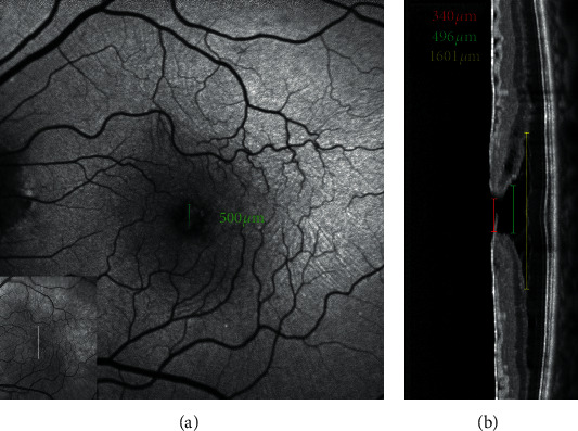
Evolution of imaging techniques has renewed interest in the diagnosis of lamellar macular hole (LMH) and greatly implemented the possibilities of gaining more detailed insights into its pathogenesis. Among noninvasive techniques, optical coherence tomography (OCT) is considered the primary examination modality to study LMHs, given its ability to image foveal structure and its widespread availability. OCT also allows to resolve the epiretinal materials associated with LMH, i.e., tractional epiretinal membranes (ERMs) and epiretinal proliferation (EP). En face OCT reconstructions are useful to confirm the foveal abnormalities shown by the eyes with LMH, whereas OCT angiography may reveal alterations of the size and shape of the foveal avascular zone and alterations of the density of the superficial and deep vascular plexuses. On slit-lamp biomicroscopy or fundus camera examination, LMH appears as a round or oval, reddish lesion at the center of the macula, slightly darker than the surrounding retina. The associated tractional ERM, causing wrinkling and glistening of the retinal surface, is usually readily appreciable, whereas EP is hardly apparent on biomicroscopy or fundus photography since the retina surface appears smooth. When imaged with blue fundus autofluorescence (B-FAF) imaging, LMHs are characterized by an increased autofluorescent signal, the intensity of which does not correlate with the thickness of the residual outer retinal tissue. Green reflectance and blue reflectance (BR) images clearly show the increased reflection and wrinkling of the retinal surface caused by tractional ERM associated with LMH. BR and multicolor imaging enable the visualization of EP associated with LMH in the form of a sharply demarcated dark area and in the form of a yellowish area surrounding the hole, respectively. Scarce data regarding invasive imaging techniques, such as fluorescein angiography, for the study of LMH are available in the literature. The aim of this review is to evaluate the contribution that each imaging modality can provide to study the morphologic characteristics of LMH.

摘要

成像技术的发展重新激发了人们对板层黄斑裂孔(LMH)诊断的兴趣,并极大地拓展了更深入了解其发病机制的可能性。在非侵入性技术中,光学相干断层扫描(OCT)因其能够对黄斑结构进行成像且广泛应用,被认为是研究LMH的主要检查方式。OCT还能够分辨与LMH相关的视网膜前物质,即牵引性视网膜前膜(ERM)和视网膜前增殖(EP)。OCT的表面重建有助于确认患有LMH的眼睛所显示的黄斑异常,而OCT血管造影可能会揭示黄斑无血管区大小和形状的改变以及浅表和深部血管丛密度的改变。在裂隙灯生物显微镜检查或眼底照相机检查中,LMH表现为黄斑中心的圆形或椭圆形红色病变,比周围视网膜略暗。通常很容易观察到相关的牵引性ERM,它会导致视网膜表面起皱和反光,而在生物显微镜检查或眼底摄影中,由于视网膜表面看起来光滑,EP很难显现。当用蓝色眼底自发荧光(B-FAF)成像时,LMH的特征是自发荧光信号增强,其强度与残留的外层视网膜组织厚度无关。绿色反射率和蓝色反射率(BR)图像清楚地显示了与LMH相关的牵引性ERM引起的视网膜表面反射增强和起皱。BR和多色成像分别能够以清晰界定的暗区形式和围绕裂孔的淡黄色区域形式显示与LMH相关的EP。关于用于研究LMH的侵入性成像技术,如荧光素血管造影的资料很少。本综述的目的是评估每种成像方式对研究LMH形态特征的贡献。

https://cdn.ncbi.nlm.nih.gov/pmc/blobs/d32b/7868145/f27b428b7bf8/joph2021-8820444.012.jpg
https://cdn.ncbi.nlm.nih.gov/pmc/blobs/d32b/7868145/00b03ac5e3d8/joph2021-8820444.001.jpg
https://cdn.ncbi.nlm.nih.gov/pmc/blobs/d32b/7868145/6cd269757ce1/joph2021-8820444.002.jpg
https://cdn.ncbi.nlm.nih.gov/pmc/blobs/d32b/7868145/d89d9923ce4f/joph2021-8820444.003.jpg
https://cdn.ncbi.nlm.nih.gov/pmc/blobs/d32b/7868145/ef1d615b23b3/joph2021-8820444.004.jpg
https://cdn.ncbi.nlm.nih.gov/pmc/blobs/d32b/7868145/f7f5661951b7/joph2021-8820444.005.jpg
https://cdn.ncbi.nlm.nih.gov/pmc/blobs/d32b/7868145/6127c4c1bb8b/joph2021-8820444.006.jpg
https://cdn.ncbi.nlm.nih.gov/pmc/blobs/d32b/7868145/57fe7cf3d016/joph2021-8820444.007.jpg
https://cdn.ncbi.nlm.nih.gov/pmc/blobs/d32b/7868145/837703ba6b19/joph2021-8820444.008.jpg
https://cdn.ncbi.nlm.nih.gov/pmc/blobs/d32b/7868145/94ab2cea3945/joph2021-8820444.009.jpg
https://cdn.ncbi.nlm.nih.gov/pmc/blobs/d32b/7868145/1f0b55102c75/joph2021-8820444.010.jpg
https://cdn.ncbi.nlm.nih.gov/pmc/blobs/d32b/7868145/e988572ac254/joph2021-8820444.011.jpg
https://cdn.ncbi.nlm.nih.gov/pmc/blobs/d32b/7868145/f27b428b7bf8/joph2021-8820444.012.jpg

相似文献

1
Multimodal Imaging of Lamellar Macular Holes.板层黄斑裂孔的多模态成像
J Ophthalmol. 2021 Jan 30;2021:8820444. doi: 10.1155/2021/8820444. eCollection 2021.
2
Epiretinal proliferation seen in association with lamellar macular holes: a distinct clinical entity.视网膜前增殖与板层黄斑裂孔相关:一种独特的临床实体。
Retina. 2014 Aug;34(8):1513-23. doi: 10.1097/IAE.0000000000000163.
3
Primary Lamellar Macular Holes: To Vit or Not to Vit.原发性板层黄斑裂孔:是否行玻璃体手术
J Clin Med. 2022 Aug 28;11(17):5046. doi: 10.3390/jcm11175046.
4
[Lamellar macular holes with hyporeflective epiretinal proliferation : OCT diagnostics and clinical course].[伴有低反射性视网膜前增殖的板层黄斑裂孔:光学相干断层扫描诊断及临床病程]
Ophthalmologe. 2017 Dec;114(12):1100-1109. doi: 10.1007/s00347-017-0597-5.
5
Long-term follow-up of fellow eye in patients with lamellar macular hole.板层黄斑裂孔患者对侧眼的长期随访
Graefes Arch Clin Exp Ophthalmol. 2017 Aug;255(8):1485-1492. doi: 10.1007/s00417-017-3652-7. Epub 2017 Apr 13.
6
Lamellar macular holes in the eyes with pathological myopia.病理性近视眼中的板层黄斑裂孔
Graefes Arch Clin Exp Ophthalmol. 2018 Jul;256(7):1281-1290. doi: 10.1007/s00417-018-3995-8. Epub 2018 May 3.
7
Assessment of Lamellar Macular Hole and Macular Pseudohole With a Combination of En Face and Radial B-scan Optical Coherence Tomography Imaging.层状黄斑裂孔和黄斑假性裂孔的评估:应用共焦和径向扫频光学相干断层扫描成像技术。
Am J Ophthalmol. 2018 Apr;188:29-40. doi: 10.1016/j.ajo.2018.01.016. Epub 2018 Jan 31.
8
The Relationship Between Blue-Fundus Autofluorescence and Optical Coherence Tomography in Eyes With Lamellar Macular Holes.板层黄斑裂孔眼的蓝眼底自发荧光与光相干断层扫描的关系。
Invest Ophthalmol Vis Sci. 2018 Jun 1;59(7):3079-3087. doi: 10.1167/iovs.18-24379.
9
Epiretinal membrane characteristics correlate with photoreceptor layer defects in lamellar macular holes and macular pseudoholes.视网膜前膜特征与板层黄斑裂孔和黄斑假性裂孔中的光感受器层缺陷相关。
Retina. 2015 Apr;35(4):727-35. doi: 10.1097/IAE.0000000000000375.
10
Optical coherence tomography angiographic findings of lamellar macular hole: comparisons between tractional and degenerative subtypes.层状黄斑裂孔的光相干断层扫描血管造影表现:牵拉型与变性型的比较。
Sci Rep. 2020 Aug 7;10(1):13331. doi: 10.1038/s41598-020-70254-0.

引用本文的文献

1
Evaluation of Optical Coherence Tomography Angiography in Degenerative and Tractional Lamellar Macular Hole.光学相干断层扫描血管造影术在退行性和牵拉性黄斑板层裂孔中的评估
J Ophthalmol. 2024 Jul 19;2024:4146294. doi: 10.1155/2024/4146294. eCollection 2024.
2
In-Depth Analysis of Preoperative OCT Markers as Prognostic Factors for Lamellar Macular Holes and Epiretinal Membrane Foveoschisis.术前 OCT 标志物对板层黄斑裂孔和视网膜前膜黄斑裂孔的预后分析。
Ophthalmol Retina. 2024 May;8(5):465-472. doi: 10.1016/j.oret.2023.10.025. Epub 2023 Nov 14.
3
Primary Lamellar Macular Holes: To Vit or Not to Vit.

本文引用的文献

1
Fluorescein Angiography Findings in Eyes With Lamellar Macular Hole and Epiretinal Membrane Foveoschisis.板层黄斑裂孔伴视网膜内界膜黄斑前膜裂孔的荧光素血管造影表现。
Invest Ophthalmol Vis Sci. 2021 Jan 4;62(1):34. doi: 10.1167/iovs.62.1.34.
2
Optical coherence tomography angiographic findings of lamellar macular hole: comparisons between tractional and degenerative subtypes.层状黄斑裂孔的光相干断层扫描血管造影表现:牵拉型与变性型的比较。
Sci Rep. 2020 Aug 7;10(1):13331. doi: 10.1038/s41598-020-70254-0.
3
Identification of epiretinal proliferation in various retinal diseases and vitreoretinal interface disorders.
原发性板层黄斑裂孔:是否行玻璃体手术
J Clin Med. 2022 Aug 28;11(17):5046. doi: 10.3390/jcm11175046.
4
Quantitative evaluation of dissociated optic nerve fibre layer (DONFL) following idiopathic macular hole surgery.特发性黄斑裂孔手术后分离的视神经纤维层 (DONFL) 的定量评估。
Eye (Lond). 2023 May;37(7):1451-1457. doi: 10.1038/s41433-022-02150-y. Epub 2022 Jul 1.
5
Macular Microvascular Modifications in Progressive Lamellar Macular Holes.进行性板层黄斑裂孔中的黄斑微血管改变
Diagnostics (Basel). 2021 Sep 19;11(9):1717. doi: 10.3390/diagnostics11091717.
各种视网膜疾病和玻璃体视网膜界面疾病中视网膜前增殖的识别。
Int J Retina Vitreous. 2020 Jul 10;6:31. doi: 10.1186/s40942-020-00233-0. eCollection 2020.
4
Optical coherence tomography-based consensus definition for lamellar macular hole.基于光学相干断层扫描的板层黄斑裂孔共识定义。
Br J Ophthalmol. 2020 Dec;104(12):1741-1747. doi: 10.1136/bjophthalmol-2019-315432. Epub 2020 Feb 27.
5
Distinctive Mechanisms and Patterns of Exudative Versus Tractional Intraretinal Cystoid Spaces as Seen With Multimodal Imaging.多模态成像所见渗出性与牵引性视网膜内囊样间隙的独特机制和模式。
Am J Ophthalmol. 2020 Apr;212:43-56. doi: 10.1016/j.ajo.2019.12.010. Epub 2019 Dec 17.
6
Foveal Abnormality associated with epiretinal Tissue of medium reflectivity and Increased blue-light fundus Autofluorescence Signal (FATIAS).与中等反射性视网膜组织和增加的蓝光眼底自发荧光信号(FATIAS)相关的中心凹异常。
Graefes Arch Clin Exp Ophthalmol. 2019 Dec;257(12):2601-2612. doi: 10.1007/s00417-019-04451-7. Epub 2019 Sep 7.
7
The role of Müller cells in tractional macular disorders: an optical coherence tomography study and physical model of mechanical force transmission.Müller 细胞在牵引性黄斑病变中的作用:光学相干断层扫描研究和机械力传递的物理模型。
Br J Ophthalmol. 2020 Apr;104(4):466-472. doi: 10.1136/bjophthalmol-2019-314245. Epub 2019 Jul 20.
8
The Relationship Between Blue-Fundus Autofluorescence and Optical Coherence Tomography in Eyes With Lamellar Macular Holes.板层黄斑裂孔眼的蓝眼底自发荧光与光相干断层扫描的关系。
Invest Ophthalmol Vis Sci. 2018 Jun 1;59(7):3079-3087. doi: 10.1167/iovs.18-24379.
9
IDENTIFICATION OF POSTERIOR SEGMENT PATHOLOGY WITH EN FACE RETINAL IMAGING USING MULTICOLOR CONFOCAL SCANNING LASER OPHTHALMOSCOPY.应用多色共焦扫描激光检眼镜的眼前节图像识别后部节段病变。
Retina. 2019 May;39(5):972-979. doi: 10.1097/IAE.0000000000002111.
10
Visualization of Lamellar Hole-Associated Epiretinal Proliferation With Blue-Reflectance Imaging.蓝光反射成像观察板层孔相关的视网膜前增殖
Retina. 2018 May;38(5):e34-e35. doi: 10.1097/IAE.0000000000002048.