Zhou Feng, Chu Linyang, Liu Xuqiang, He Zihao, Han Xuequan, Yan Mengning, Qu Xinhua, Li Xiaofeng, Yu Zhifeng
Shanghai Key Laboratory of Orthopaedic Implants, Department of Orthopaedic Surgery, Shanghai Ninth People's Hospital, Shanghai Jiao Tong University School of Medicine, Shanghai, China.
Department of Orthopaedic Surgery, First Affiliated Hospital of Soochow University, Suzhou, China.
Front Med (Lausanne). 2021 Feb 2;8:617200. doi: 10.3389/fmed.2021.617200. eCollection 2021.
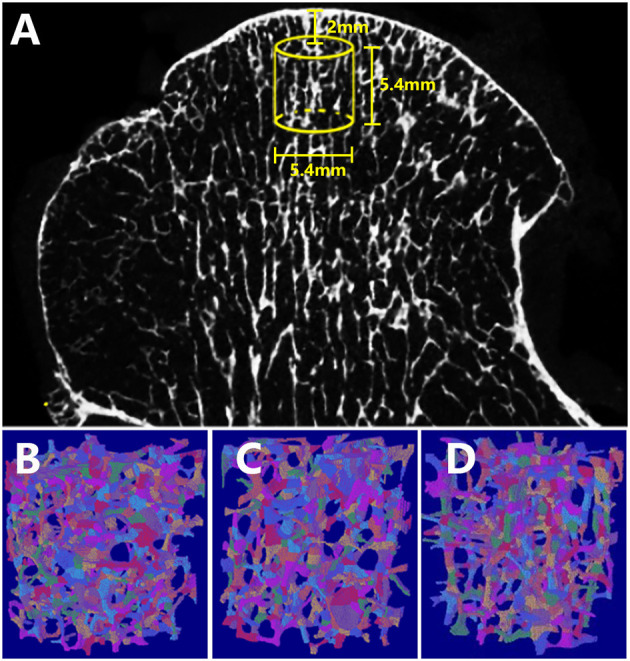
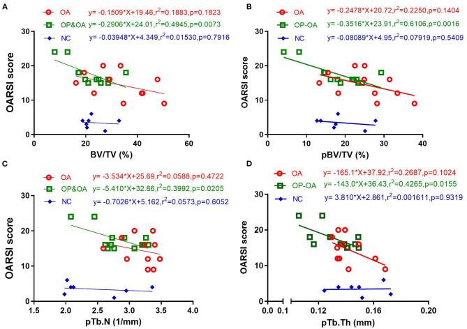
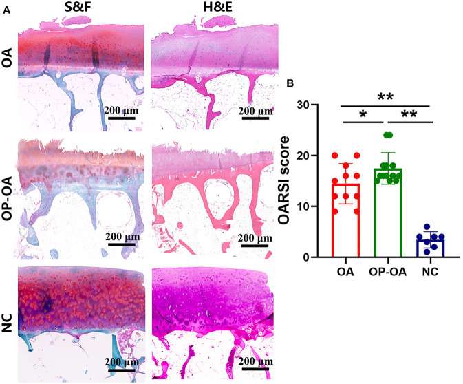
Osteoporotic osteoarthritis (OP-OA) is a specific type of OA. In this study, we aimed to assess the subchondral plate and rod microstructural differences between OA and OP-OA patients by using an individual trabeculae segmentation (ITS) system and to analyze the relationships between subchondral microstructures and cartilage damage in OA and OP-OA patients. Overall, 31 femoral heads were included in this study, which included 11 samples with OA and 13 samples with OP-OA; the normal control (NC) group contained 7 healthy femoral heads. ITS was performed to segment the subchondral trabecular bone into plate and rod trabeculae based on microcomputed tomography (micro-CT) images. We compared the plate and rod trabeculae of the subchondral trabecular bone between OA and OP-OA patients. The Osteoarthritis Research Society International (OARSI) score was employed to evaluate cartilage damage based on histological observations. Pearson's correlation coefficient and linear regression analysis were applied to analyze the relationships between subchondral microstructures and articular cartilage damage. Results showed that several microstructural parameters, including bone volume fraction (BV/TV), plate bone volume fraction (pBV/TV), rod bone volume fraction (rBV/TV), plate trabecular number (pTb.N), rod trabecular number (rTb.N), junction density between rod and plate (R-P Junc.D), and junction density between plate and plate (P-P Junc.D), were significantly decreased in patients with OP-OA compared with those in patients with OA ( < 0.05). Histological observations indicated that cartilage damage was more serious in patients with OP-OA than that in patients with OA ( < 0.05). Moreover, BV/TV, pBV/TV, pTb.N, and pTb.Th were significantly related to the OARSI score in both OA and OP-OA patients. These results indicated that there were differences in the subchondral rod and plate trabeculae between OA and OP-OA patients. Subchondral decreased plate trabeculae (pBV/TV, pTb.N, and pTb.Th) might account for cartilage damage in the progression of OP-OA. This study provided new insights to research OA when it is combined with OP.
骨质疏松性骨关节炎(OP - OA)是骨关节炎的一种特殊类型。在本研究中,我们旨在通过使用个体小梁分割(ITS)系统评估骨关节炎患者与骨质疏松性骨关节炎患者的软骨下骨板和骨小梁的微观结构差异,并分析骨关节炎和骨质疏松性骨关节炎患者软骨下微观结构与软骨损伤之间的关系。本研究共纳入31个股骨头,其中骨关节炎样本11例,骨质疏松性骨关节炎样本13例;正常对照组(NC)包含7个健康股骨头。基于微计算机断层扫描(micro - CT)图像,运用ITS将软骨下骨小梁分割为骨板和骨小梁。我们比较了骨关节炎患者与骨质疏松性骨关节炎患者软骨下骨小梁的骨板和骨小梁情况。采用国际骨关节炎研究学会(OARSI)评分,根据组织学观察评估软骨损伤。应用Pearson相关系数和线性回归分析来分析软骨下微观结构与关节软骨损伤之间的关系。结果显示,与骨关节炎患者相比,骨质疏松性骨关节炎患者的几个微观结构参数,包括骨体积分数(BV/TV)、骨板骨体积分数(pBV/TV)、骨小梁骨体积分数(rBV/TV)、骨板小梁数量(pTb.N)、骨小梁小梁数量(rTb.N)、骨小梁与骨板之间的连接密度(R - P Junc.D)以及骨板与骨板之间的连接密度(P - P Junc.D)均显著降低(<0.05)。组织学观察表明,骨质疏松性骨关节炎患者的软骨损伤比骨关节炎患者更严重(<0.05)。此外,在骨关节炎和骨质疏松性骨关节炎患者中,BV/TV、pBV/TV、pTb.N和pTb.Th均与OARSI评分显著相关。这些结果表明,骨关节炎患者与骨质疏松性骨关节炎患者的软骨下骨小梁和骨板存在差异。软骨下骨板小梁减少(pBV/TV、pTb.N和pTb.Th)可能是骨质疏松性骨关节炎进展过程中软骨损伤的原因。本研究为研究骨关节炎合并骨质疏松提供了新的见解。