Chu Linyang, He Zihao, Qu Xinhua, Liu Xuqiang, Zhang Weituo, Zhang Shuo, Han Xuequan, Yan Mengning, Xu Qi, Zhang Shuhong, Shang Xifu, Yu Zhifeng
Shanghai Key Laboratory of Orthopedic Implants, Department of Orthopedic Surgery, Shanghai Ninth People's Hospital, Shanghai Jiao Tong University School of Medicine, Shanghai, PR China.
Department of Bone and Joint Surgery, Renji Hospital, Shanghai Jiaotong University School of Medicine, Shanghai, 200011, PR China.
J Orthop Translat. 2019 Sep 27;22:50-57. doi: 10.1016/j.jot.2019.09.001. eCollection 2020 May.
Developmental dysplasia of the hip (DDH) is recognized as a frequent cause of secondary osteoarthritis (OA). The purpose in this study was to compare structural and biomechanical properties of subchondral trabecular bone and its relationship with cartilage damage between patients with DDH and patients with primary hip OA.
Forty-three femoral head specimens obtained from patients who underwent total hip arthroplasty [DDH, n = 17; primary OA, n = 16; and normal control (NC), n = 10] were scanned by microcomputed tomography and analyzed by individual trabecula segmentation to obtain the microstructural types of subchondral trabecular bone. The biomechanical properties were analyzed by micro-finite element analysis, and cartilage damage was evaluated by histology. The linear regression analysis was used to indicate the association between microstructures, biomechanical property, and articular cartilage.
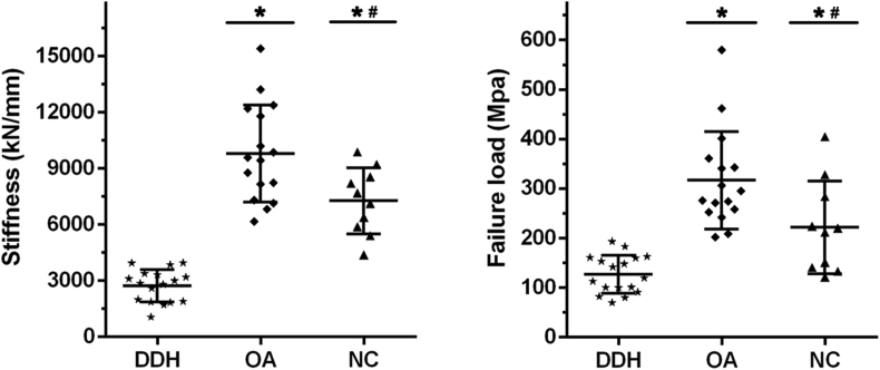
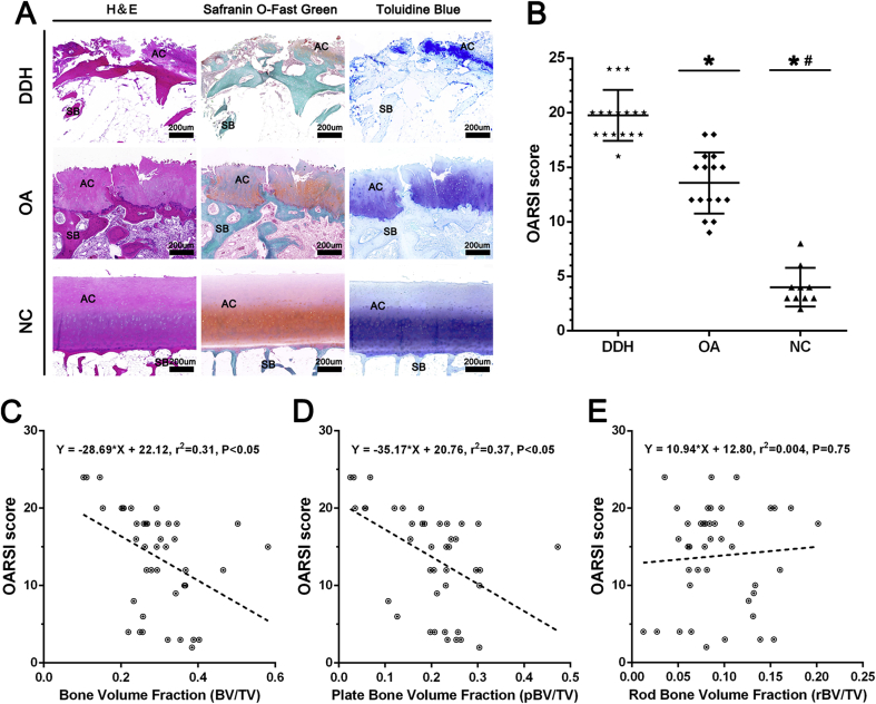
The DDH group showed the lowest total bone volume fractions (BV/TV) and plate BV/TV in the three groups (p < 0.05). There were also different discrepancies between the three groups in plate/rod trabecular number, plate/rod trabecular thickness, trabecular plate surface area/trabecular rod length, and junction density with different modes (plate-plate, rod-rod, and plate-rod junction density). The micro-finite element analysis, histology, and linear regression revealed that the subchondral trabecular bone in the DDH group had inferior biomechanical properties and cartilage damage of patients with DDH was more serious with different subchondral trabecular bone microstructures.
Our findings detected deteriorating subchondral trabecular bone microstructures in patients with DDH. The mass and type of subchondral trabecular bone play a key role in mechanical properties in DDH, which might be related to cartilage damage.
Our findings suggested that changes of subchondral trabecular bone play a critical role in DDH progression and that the improvement on subchondral trabecular bone may be a sensitive and promising way in treatment of DDH.
发育性髋关节发育不良(DDH)被认为是继发性骨关节炎(OA)的常见病因。本研究旨在比较DDH患者与原发性髋关节OA患者的软骨下小梁骨的结构和生物力学特性及其与软骨损伤的关系。
对43例接受全髋关节置换术患者的股骨头标本进行扫描[DDH组,n = 17;原发性OA组,n = 16;正常对照组(NC),n = 10],采用微型计算机断层扫描,并通过个体小梁分割进行分析,以获得软骨下小梁骨的微观结构类型。通过微有限元分析评估生物力学特性,通过组织学评估软骨损伤。采用线性回归分析来表明微观结构、生物力学特性和关节软骨之间的关联。
DDH组的总骨体积分数(BV/TV)和板层BV/TV在三组中最低(p < 0.05)。三组在板/杆小梁数量、板/杆小梁厚度、小梁板表面积/小梁杆长度以及不同模式(板-板、杆-杆和板-杆连接密度)的连接密度方面也存在不同差异。微有限元分析、组织学和线性回归显示,DDH组的软骨下小梁骨生物力学特性较差,且不同软骨下小梁骨微观结构的DDH患者软骨损伤更严重。
我们的研究结果发现DDH患者的软骨下小梁骨微观结构恶化。软骨下小梁骨的质量和类型在DDH的力学性能中起关键作用,这可能与软骨损伤有关。
我们的研究结果表明,软骨下小梁骨的变化在DDH进展中起关键作用,改善软骨下小梁骨可能是治疗DDH的一种敏感且有前景的方法。