Department of Functional Neuroanatomy, Institute for Anatomy and Cell Biology, Heidelberg University, Im Neuenheimer Feld 307, 69120, Heidelberg, Germany.
Translational Imaging Research Group, Central Institute of Mental Health, Medical Faculty of Mannheim, Heidelberg University, J5, 68159, Mannheim, Germany.
Sci Rep. 2021 Feb 19;11(1):4234. doi: 10.1038/s41598-021-83491-8.
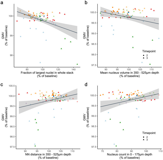
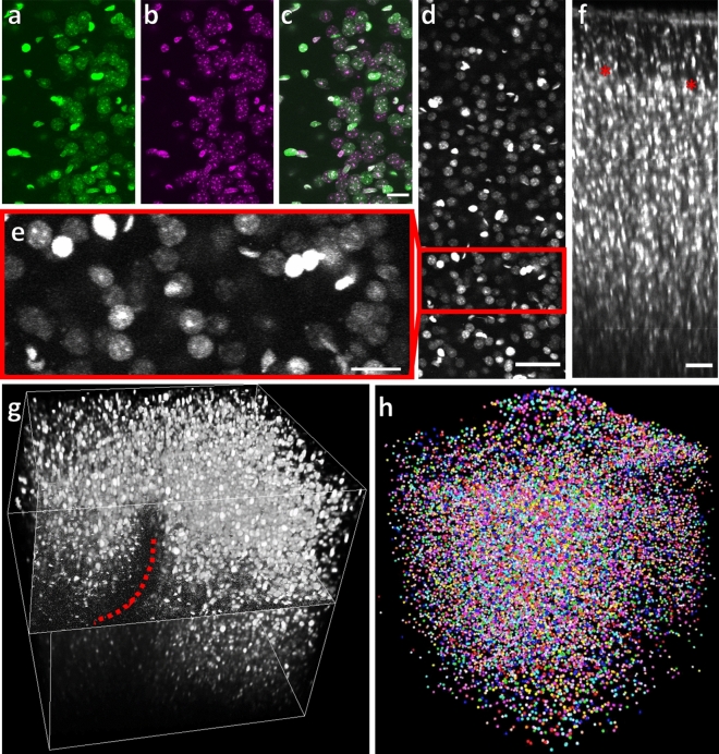
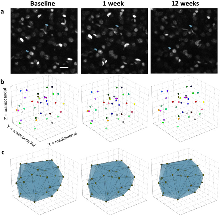
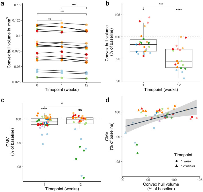
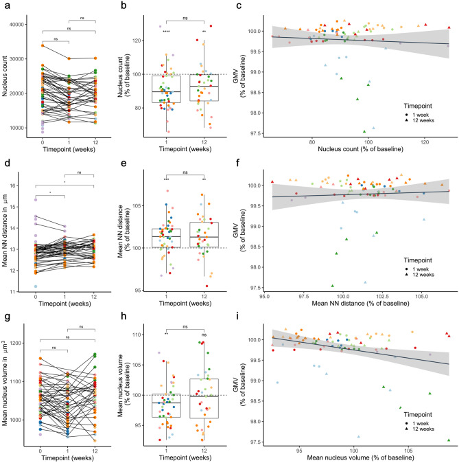
Magnetic resonance imaging (MRI) of the brain combined with voxel-based morphometry (VBM) revealed changes in gray matter volume (GMV) in various disorders. However, the cellular basis of GMV changes has remained largely unclear. We correlated changes in GMV with cellular metrics by imaging mice with MRI and two-photon in vivo microscopy at three time points within 12 weeks, taking advantage of age-dependent changes in brain structure. Imaging fluorescent cell nuclei allowed inferences on (i) physical tissue volume as determined from reference spaces outlined by nuclei, (ii) cell density, (iii) the extent of cell clustering, and (iv) the volume of cell nuclei. Our data indicate that physical tissue volume alterations only account for 13.0% of the variance in GMV change. However, when including comprehensive measurements of nucleus volume and cell density, 35.6% of the GMV variance could be explained, highlighting the influence of distinct cellular mechanisms on VBM results.
脑磁共振成像(MRI)结合体素形态计量学(VBM)显示,在各种疾病中,灰质体积(GMV)发生了变化。然而,GMV 变化的细胞基础在很大程度上仍不清楚。我们通过 MRI 和双光子在 12 周内的三个时间点对小鼠进行活体成像,利用脑结构的年龄依赖性变化,将 GMV 的变化与细胞指标相关联。对荧光细胞核成像可推断出:(i)由细胞核界定的参考空间中的实际组织体积;(ii)细胞密度;(iii)细胞聚类程度;以及(iv)细胞核体积。我们的数据表明,实际组织体积的改变仅占 GMV 变化的 13.0%。然而,当包括细胞核体积和细胞密度的综合测量时,可解释 35.6%的 GMV 方差,突出了不同细胞机制对 VBM 结果的影响。