Wu Zhanxiong, Peng Yun, Hong Ming, Zhang Yingchun
School of Electronic Information, Hangzhou Dianzi University, Hangzhou, China.
Department of Biomedical Engineering, University of Houston, Houston, TX, United States.
Front Aging Neurosci. 2021 Feb 3;13:593898. doi: 10.3389/fnagi.2021.593898. eCollection 2021.
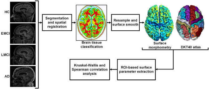
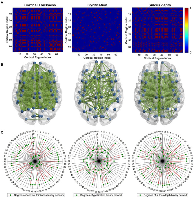
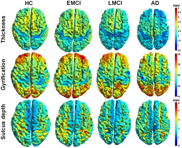
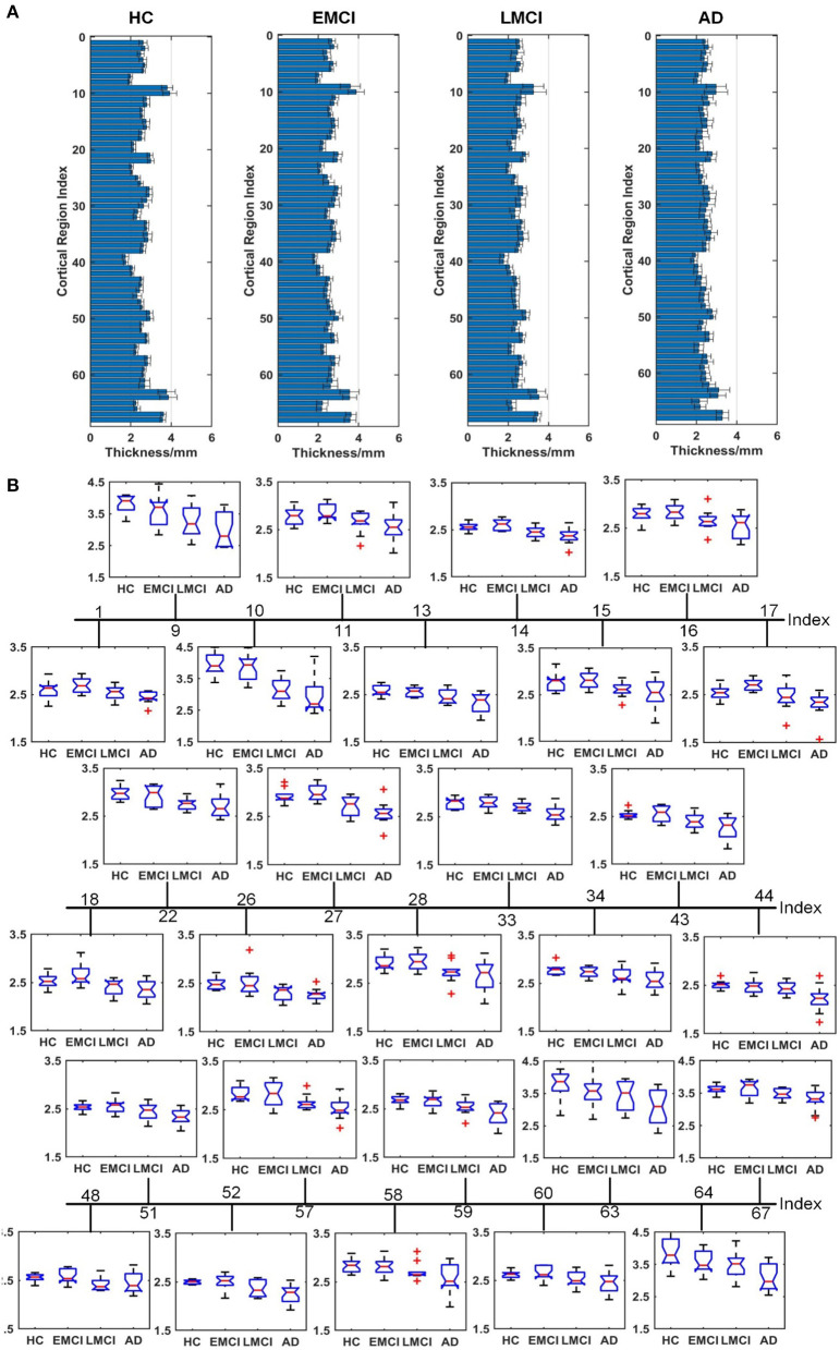
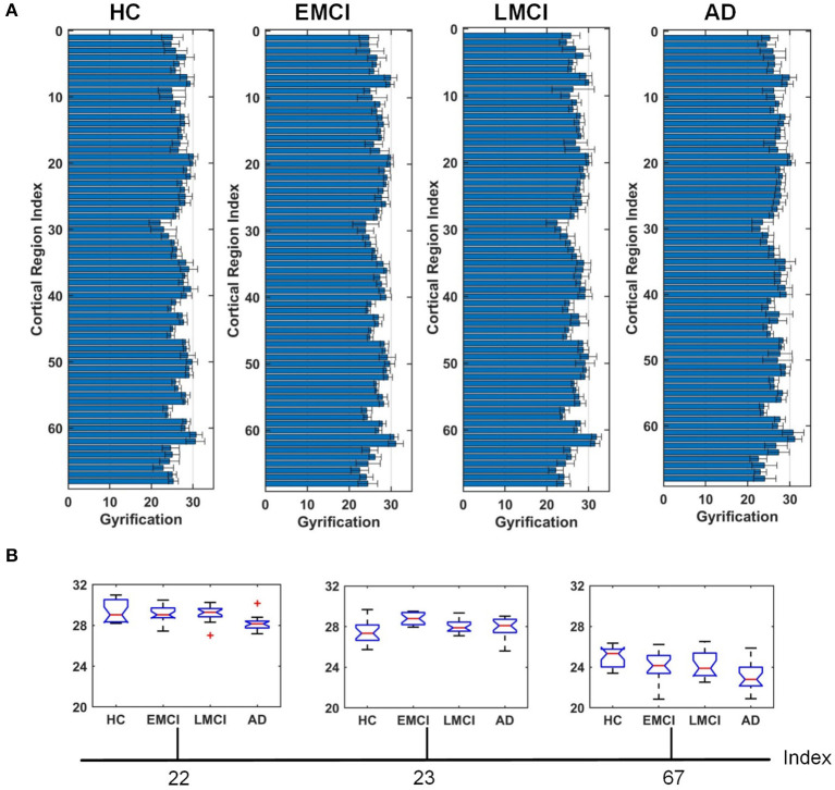
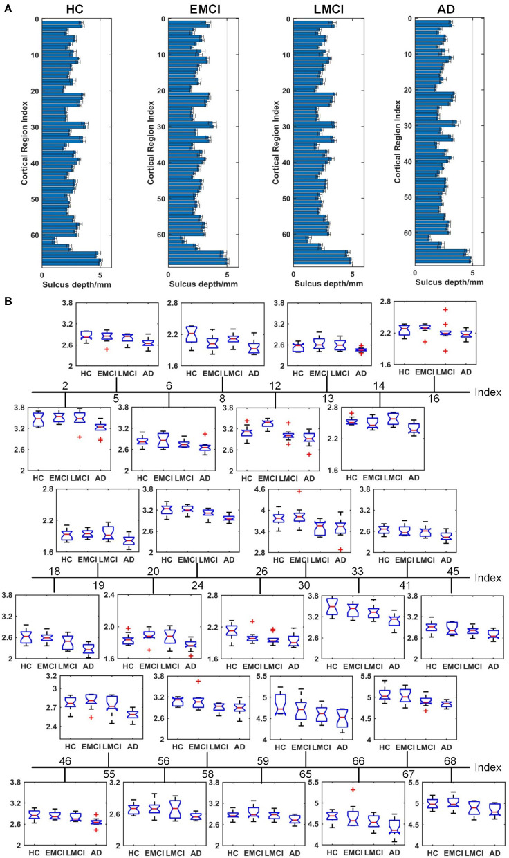
Accurate detection of the regions of Alzheimer's disease (AD) lesions is critical for early intervention to effectively slow down the progression of the disease. Although gray matter volumetric abnormalities are commonly detected in patients with mild cognition impairment (MCI) and patients with AD, the gray matter surface-based deterioration pattern associated with the progression of the disease from MCI to AD stages is largely unknown. To identify group differences in gray matter surface morphometry, including cortical thickness, the gyrification index (GI), and the sulcus depth, 80 subjects from the Alzheimer's Disease Neuroimaging Initiative (ADNI) database were split into healthy controls (HCs; = 20), early MCIs (EMCI; = 20), late MCIs (LMCI; = 20), and ADs ( = 20). Regions-of-interest (ROI)-based surface morphometry was subsequently studied and compared across the four stage groups to characterize the gray matter deterioration during AD progression. Co-alteration patterns (Spearman's correlation coefficient) across the whole brain were also examined. Results showed that patients with MCI and AD exhibited a significant reduction in cortical thickness ( < 0.001) mainly in the cingulate region (four subregions) and in the temporal (thirteen subregions), parietal (five subregions), and frontal (six subregions) lobes compared to HCs. The sulcus depth of the eight temporal, four frontal, four occipital, and eight parietal subregions were also significantly affected ( < 0.001) by the progression of AD. The GI was shown to be insensitive to AD progression (only three subregions were detected with a significant difference, < 0.001). Moreover, Spearman's correlation analysis confirmed that the co-alteration pattern of the cortical thickness and sulcus depth indices is predominant during AD progression. The findings highlight the relevance between gray matter surface morphometry and the stages of AD, laying the foundation for tracking of AD progression. The co-alteration pattern of surface-based morphometry would improve the researchers' knowledge of the underlying pathologic mechanisms in AD.
准确检测阿尔茨海默病(AD)病变区域对于早期干预以有效减缓疾病进展至关重要。尽管在轻度认知障碍(MCI)患者和AD患者中通常能检测到灰质体积异常,但与疾病从MCI进展到AD阶段相关的基于灰质表面的恶化模式在很大程度上尚不清楚。为了确定灰质表面形态测量的组间差异,包括皮质厚度、脑回化指数(GI)和脑沟深度,将阿尔茨海默病神经成像计划(ADNI)数据库中的80名受试者分为健康对照组(HCs;n = 20)、早期MCI(EMCI;n = 20)、晚期MCI(LMCI;n = 20)和AD组(n = 20)。随后研究并比较了四个阶段组基于感兴趣区域(ROI)的表面形态测量,以表征AD进展过程中的灰质恶化情况。还检查了全脑的共同改变模式(斯皮尔曼相关系数)。结果显示,与HCs相比,MCI和AD患者的皮质厚度显著降低(P < 0.001),主要集中在扣带区域(四个亚区域)以及颞叶(十三个亚区域)、顶叶(五个亚区域)和额叶(六个亚区域)。AD进展也显著影响了八个颞叶、四个额叶、四个枕叶和八个顶叶亚区域的脑沟深度(P < 0.001)。结果表明GI对AD进展不敏感(仅检测到三个亚区域有显著差异,P < 0.001)。此外,斯皮尔曼相关分析证实,在AD进展过程中,皮质厚度和脑沟深度指数的共同改变模式占主导。这些发现突出了灰质表面形态测量与AD阶段之间的相关性,为追踪AD进展奠定了基础。基于表面的形态测量的共同改变模式将提高研究人员对AD潜在病理机制的认识。