• 文献检索
  • 文档翻译
  • 深度研究
  • 学术资讯
  • Suppr Zotero 插件Zotero 插件
  • 邀请有礼
  • 套餐&价格
  • 历史记录
应用&插件
Suppr Zotero 插件Zotero 插件浏览器插件Mac 客户端Windows 客户端微信小程序
定价
高级版会员购买积分包购买API积分包
服务
文献检索文档翻译深度研究API 文档MCP 服务
关于我们
关于 Suppr公司介绍联系我们用户协议隐私条款
关注我们

Suppr 超能文献

核心技术专利:CN118964589B侵权必究
粤ICP备2023148730 号-1Suppr @ 2026

文献检索

告别复杂PubMed语法,用中文像聊天一样搜索,搜遍4000万医学文献。AI智能推荐,让科研检索更轻松。

立即免费搜索

文件翻译

保留排版,准确专业,支持PDF/Word/PPT等文件格式,支持 12+语言互译。

免费翻译文档

深度研究

AI帮你快速写综述,25分钟生成高质量综述,智能提取关键信息,辅助科研写作。

立即免费体验

术前 [F]FDG-PET/CT 对外阴癌淋巴结分期的诊断性能:一项大型单中心研究。

Diagnostic performance of preoperative [F]FDG-PET/CT for lymph node staging in vulvar cancer: a large single-centre study.

机构信息

Section of Nuclear Medicine, University Department of Radiological Sciences and Haematology, Università Cattolica del Sacro Cuore, Largo A. Gemelli, 8, 00168, Rome, Italy.

Unit of Nuclear Medicine, Fondazione Policlinico Universitario A. Gemelli IRCCS, Rome, Italy.

出版信息

Eur J Nucl Med Mol Imaging. 2021 Sep;48(10):3303-3314. doi: 10.1007/s00259-021-05257-8. Epub 2021 Feb 23.

DOI:10.1007/s00259-021-05257-8
PMID:33619601
原文链接:https://pmc.ncbi.nlm.nih.gov/articles/PMC8426310/
Abstract

PURPOSE

This retrospective study aimed to assess the diagnostic performance of preoperative [F]FDG-PET/CT in predicting the groin and pelvic lymph node (LN) status in a large single-centre series of vulvar cancer patients.

METHODS

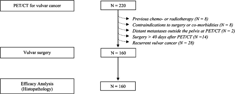
Between January 2013 and October 2018, among all consecutive women with proven vulvar cancer submitted to [F]FDG-PET/CT, 160 patients were included. LNs were analysed by two qualitative methods assessing PET information (defined as visual assessment) and a combination of PET and low-dose CT information (defined as overall assessment), respectively, as well as semi-quantitative analysis (LN-SUV). Sensitivity, specificity, accuracy, positive and negative predictive values (PPV and NPV) in predicting the groin and pelvic LN status were calculated in the overall study population; a subset analysis of groin parameters in clinically/ultrasonography negative patients was also performed. Histopathology was the reference standard.

RESULTS

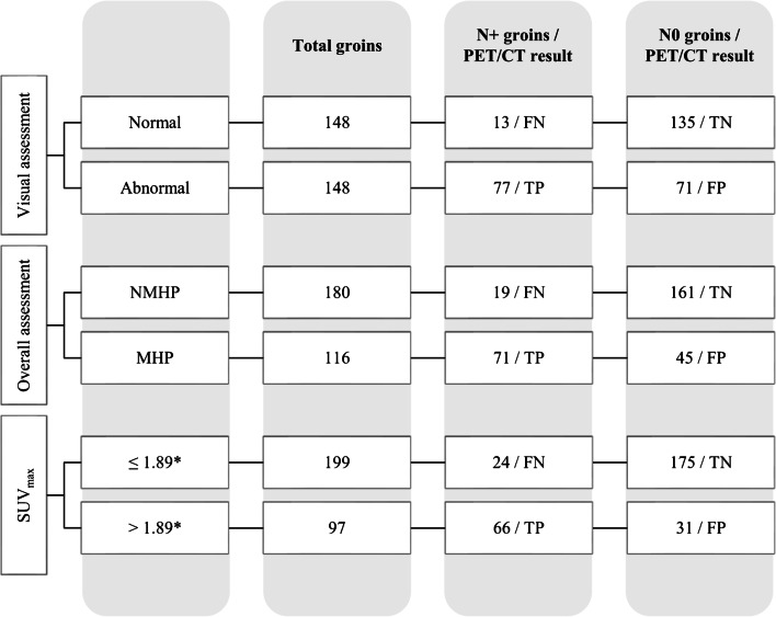
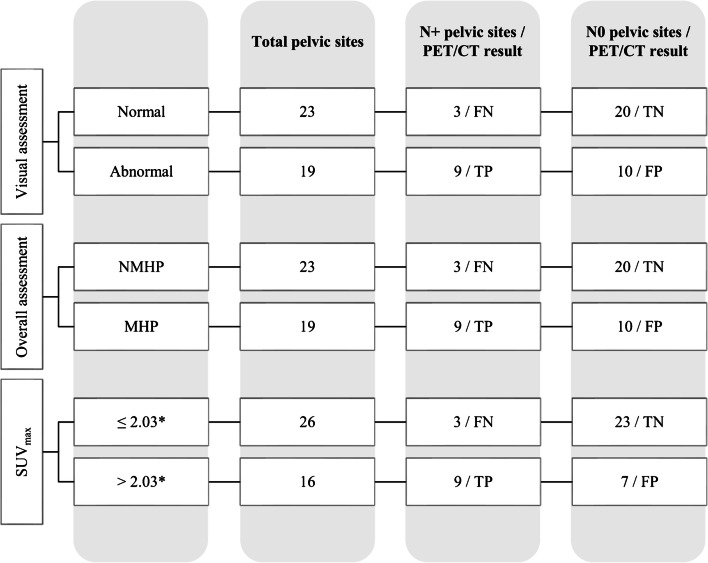
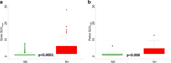
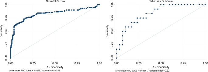
All patients underwent vulvar and inguinofemoral LN surgery, and 35 pelvic LN surgery. Overall, 338 LN sites (296 groins and 42 pelvic sites) were histologically examined with 30.4% prevalence of metastatic groins and 28.6% for metastatic pelvic sites. In the overall study population, sensitivity (95% confidence interval, CI), specificity (95% CI), accuracy (95% CI), PPV (95% CI) and NPV (95% CI) at the groin level were 85.6% (78.3-92.8), 65.5% (59.0-72.0), 71.6% (66.5-76.8), 52.0% (44.0-60.1) and 91.2% (86.7-95.8) for visual assessment; 78.9% (70.5-87.3), 78.2% (72.5-83.8), 78.4% (73.7-83.1), 61.2% (52.3-70.1) and 89.4% (85.0-93.9) for overall assessment; and 73.3% (64.2-82.5), 85.0% (80.1-89.8), 81.4% (77.0-85.8), 68.0% (58.8-77.3) and 87.9% (83.4-92.5) for semi-quantitative analysis (SUV cut-off value 1.89 achieved by ROC analysis). Similar results were observed in the pelvis-based analysis.

CONCLUSION

In this large single-centre series of vulvar cancer patients, [F]FDG-PET/CT showed good values of sensitivity and NPV in discriminating metastatic from non-metastatic LNs. In routine clinical practice, qualitative analysis is a reliable interpretative criterion making unnecessary commonly used semi-quantitative methods such as SUV.

摘要

目的

本回顾性研究旨在评估术前 [F]FDG-PET/CT 在预测大样本单一中心外阴癌患者腹股沟和盆腔淋巴结(LN)状态方面的诊断性能。

方法

2013 年 1 月至 2018 年 10 月,在所有经证实患有外阴癌并接受 [F]FDG-PET/CT 的连续女性患者中,纳入 160 例患者。通过两种定性方法(定义为视觉评估)和一种 PET 和低剂量 CT 信息相结合的方法(定义为总体评估)分别对 LN 进行分析,以及半定量分析(LN-SUV)。在整个研究人群中计算了预测腹股沟和盆腔 LN 状态的灵敏度、特异性、准确性、阳性预测值(PPV)和阴性预测值(NPV);还对临床/超声阴性患者的腹股沟参数进行了亚组分析。组织病理学是参考标准。

结果

所有患者均接受了外阴和腹股沟股淋巴结手术,35 例接受了盆腔淋巴结手术。总共检查了 338 个 LN 部位(296 个腹股沟和 42 个盆腔部位),腹股沟转移性淋巴结的患病率为 30.4%,盆腔转移性淋巴结的患病率为 28.6%。在整个研究人群中,腹股沟水平的灵敏度(95%置信区间,CI)、特异性(95%CI)、准确性(95%CI)、PPV(95%CI)和 NPV(95%CI)分别为 85.6%(78.3-92.8)、65.5%(59.0-72.0)、71.6%(66.5-76.8)、52.0%(44.0-60.1)和 91.2%(86.7-95.8);78.9%(70.5-87.3)、78.2%(72.5-83.8)、78.4%(73.7-83.1)、61.2%(52.3-70.1)和 89.4%(85.0-93.9);73.3%(64.2-82.5)、85.0%(80.1-89.8)、81.4%(77.0-85.8)、68.0%(58.8-77.3)和 87.9%(83.4-92.5)。半定量分析(通过 ROC 分析获得的 SUV 截断值为 1.89)也得到了类似的结果。

结论

在这项大型单一中心外阴癌患者研究中,[F]FDG-PET/CT 在区分转移性和非转移性淋巴结方面具有良好的灵敏度和 NPV 值。在常规临床实践中,定性分析是一种可靠的解释标准,不需要常用的半定量方法(如 SUV)。

https://cdn.ncbi.nlm.nih.gov/pmc/blobs/e876/8426310/6278aab06e6e/259_2021_5257_Fig6_HTML.jpg
https://cdn.ncbi.nlm.nih.gov/pmc/blobs/e876/8426310/97526aed74d6/259_2021_5257_Fig1_HTML.jpg
https://cdn.ncbi.nlm.nih.gov/pmc/blobs/e876/8426310/5eab1251ac57/259_2021_5257_Fig2_HTML.jpg
https://cdn.ncbi.nlm.nih.gov/pmc/blobs/e876/8426310/848881ac02c6/259_2021_5257_Fig3_HTML.jpg
https://cdn.ncbi.nlm.nih.gov/pmc/blobs/e876/8426310/5911453f3207/259_2021_5257_Fig4_HTML.jpg
https://cdn.ncbi.nlm.nih.gov/pmc/blobs/e876/8426310/8dec38a23fd5/259_2021_5257_Fig5_HTML.jpg
https://cdn.ncbi.nlm.nih.gov/pmc/blobs/e876/8426310/6278aab06e6e/259_2021_5257_Fig6_HTML.jpg

相似文献

1
Diagnostic performance of preoperative [F]FDG-PET/CT for lymph node staging in vulvar cancer: a large single-centre study.术前 [F]FDG-PET/CT 对外阴癌淋巴结分期的诊断性能:一项大型单中心研究。
Eur J Nucl Med Mol Imaging. 2021 Sep;48(10):3303-3314. doi: 10.1007/s00259-021-05257-8. Epub 2021 Feb 23.
2
Evaluation of Dual-Timepoint F-FDG PET/CT Imaging for Lymph Node Staging in Vulvar Cancer.双时相F-FDG PET/CT成像在外阴癌淋巴结分期中的评估
J Nucl Med. 2017 Dec;58(12):1913-1918. doi: 10.2967/jnumed.117.194332. Epub 2017 May 25.
3
18F-FDG PET/CT in preoperative staging of vulvar cancer patients: is it really effective?18F-FDG PET/CT在外阴癌患者术前分期中的应用:真的有效吗?
Medicine (Baltimore). 2017 Sep;96(38):e7943. doi: 10.1097/MD.0000000000007943.
4
Groin sentinel node biopsy and F-FDG PET/CT-supported preoperative lymph node assessment in cN0 patients with vulvar cancer currently unfit for minimally invasive inguinal surgery: The GroSNaPET study.腹股沟前哨淋巴结活检及F-FDG PET/CT支持的术前淋巴结评估在目前不适合微创腹股沟手术的cN0期外阴癌患者中的应用:GroSNaPET研究
Eur J Surg Oncol. 2017 Sep;43(9):1776-1783. doi: 10.1016/j.ejso.2017.06.018. Epub 2017 Jul 16.
5
Accuracy of F-18 FDG PET/CT with optimal cut-offs of maximum standardized uptake value according to size for diagnosis of regional lymph node metastasis in patients with rectal cancer.根据大小设定 FDG PET/CT 最大标准化摄取值最佳截断值对直肠癌患者区域淋巴结转移的诊断准确性。
Cancer Imaging. 2018 Sep 14;18(1):32. doi: 10.1186/s40644-018-0165-5.
6
F-18 fluoro-D-glucose (FDG)-positron emission tomography (PET)/computed tomography (CT) in planning of surgery and sentinel lymph node screening in vulvar cancers.F-18氟代-D-葡萄糖(FDG)正电子发射断层显像(PET)/计算机断层扫描(CT)在外阴癌手术规划及前哨淋巴结筛查中的应用
Arch Gynecol Obstet. 2016 Jun;293(6):1319-24. doi: 10.1007/s00404-015-3927-3. Epub 2015 Oct 23.
7
Ultrasound morphometric and cytologic preoperative assessment of inguinal lymph-node status in women with vulvar cancer: MorphoNode study.外阴癌患者腹股沟淋巴结状态的超声形态和细胞学术前评估:MorphoNode 研究。
Ultrasound Obstet Gynecol. 2020 Mar;55(3):401-410. doi: 10.1002/uog.20378.
8
Use of 18F-fluoro-2-deoxy-d-glucose (18F-FDG) PET/CT for lymph node assessment before radical cystectomy in bladder cancer patients.在膀胱癌患者根治性膀胱切除术前使用 18F-氟代-2-脱氧-D-葡萄糖(18F-FDG)PET/CT 进行淋巴结评估。
BJU Int. 2024 Oct;134(4):636-643. doi: 10.1111/bju.16363. Epub 2024 Apr 15.
9
Whole-body [18F]fluoro-2-deoxyglucose positron emission tomography scan as combined PET-CT staging prior to planned radical vulvectomy and inguinofemoral lymphadenectomy for squamous vulvar cancer: a correlation with groin node metastasis.对于计划行根治性外阴切除术及腹股沟股淋巴结清扫术的鳞状外阴癌患者,在术前进行全身[18F]氟-2-脱氧葡萄糖正电子发射断层扫描作为PET-CT联合分期:与腹股沟淋巴结转移的相关性
Eur J Gynaecol Oncol. 2014;35(3):230-5.
10
Integrated analysis of F-FDG PET/CT improves preoperative lymph node staging for patients with invasive bladder cancer.F-FDG PET/CT 融合分析提高浸润性膀胱癌患者术前淋巴结分期的准确性。
Eur Radiol. 2019 Aug;29(8):4286-4293. doi: 10.1007/s00330-018-5959-0. Epub 2019 Jan 21.

引用本文的文献

1
Random Forest-Based Detection of Metastases in Clinically Scanned Lymph Nodes Using Quantitative Ultrasound Imaging.基于随机森林的定量超声成像技术在临床扫描淋巴结转移检测中的应用
Ultrasound Med Biol. 2025 Sep;51(9):1439-1446. doi: 10.1016/j.ultrasmedbio.2025.05.014. Epub 2025 Jun 19.
2
Management of Patients with Vulvar Cancers: A Systematic Comparison of International Guidelines (NCCN-ASCO-ESGO-BGCS-IGCS-FIGO-French Guidelines-RCOG).外阴癌患者的管理:国际指南(美国国立综合癌症网络-美国临床肿瘤学会-欧洲妇科肿瘤学会-巴西妇科肿瘤学会-国际妇产科联盟-法国指南-英国皇家妇产科医师学院)的系统比较
Cancers (Basel). 2025 Jan 8;17(2):186. doi: 10.3390/cancers17020186.
3

本文引用的文献

1
18F-FDG PET and 18F-FDG PET/CT in Vulvar Cancer: A Systematic Review and Meta-analysis.18F-FDG PET 和 18F-FDG PET/CT 在外阴癌中的应用:系统评价和荟萃分析。
Clin Nucl Med. 2021 Feb 1;46(2):125-132. doi: 10.1097/RLU.0000000000003411.
2
Radiologic Assessment of Groin Lymph Nodes in Pelvic Malignancies.盆腔恶性肿瘤腹股沟淋巴结的放射学评估。
Int J Gynecol Cancer. 2020 Jul;30(7):947-953. doi: 10.1136/ijgc-2020-001363. Epub 2020 Jun 2.
3
Lymphoscintigraphy and sentinel lymph node biopsy in vulvar carcinoma: update from a European expert panel.
Investigation of the distribution of inguinal lymph nodes and delineation of the inguinal clinical target volume using F-FDG PET/CT.
使用 F-FDG PET/CT 研究腹股沟淋巴结的分布并勾画腹股沟临床靶区。
BMC Cancer. 2024 Oct 10;24(1):1254. doi: 10.1186/s12885-024-13015-w.
4
Imaging in Vulval Cancer.外阴癌的影像学检查
Cancers (Basel). 2024 Jun 19;16(12):2269. doi: 10.3390/cancers16122269.
5
Current Preoperative Management of Vulvar Squamous Cell Carcinoma: An Overview.外阴鳞状细胞癌的当前术前管理:概述
Cancers (Basel). 2024 May 11;16(10):1846. doi: 10.3390/cancers16101846.
6
Application of PET/MRI in Gynecologic Malignancies.PET/MRI在妇科恶性肿瘤中的应用。
Cancers (Basel). 2024 Apr 12;16(8):1478. doi: 10.3390/cancers16081478.
7
Staging by imaging in gynecologic cancer and the role of ultrasound: an update of European joint consensus statements.妇科癌症的影像学分期和超声的作用:欧洲联合共识声明的更新。
Int J Gynecol Cancer. 2024 Mar 4;34(3):363-378. doi: 10.1136/ijgc-2023-004609.
8
Intelligent ultrafast total-body PET for sedation-free pediatric [F]FDG imaging.智能超快速全身 PET 用于无镇静儿科 [F]FDG 成像。
Eur J Nucl Med Mol Imaging. 2024 Jul;51(8):2353-2366. doi: 10.1007/s00259-024-06649-2. Epub 2024 Feb 22.
9
The Utility of F-FDG-PET-CT Metabolic Parameters in Evaluating the Primary Tumor Aggressiveness and Lymph Node Metastasis of Nasopharyngeal Carcinoma.F-FDG-PET-CT代谢参数在评估鼻咽癌原发肿瘤侵袭性和淋巴结转移中的应用
Clin Med Insights Oncol. 2024 Feb 5;18:11795549231225419. doi: 10.1177/11795549231225419. eCollection 2024.
10
Molecular image-guided surgery in gynaecological cancer: where do we stand?妇科恶性肿瘤的分子影像引导手术:我们处于什么位置?
Eur J Nucl Med Mol Imaging. 2024 Aug;51(10):3026-3039. doi: 10.1007/s00259-024-06604-1. Epub 2024 Jan 18.
外阴癌的淋巴闪烁显像和前哨淋巴结活检:欧洲专家小组的最新进展
Eur J Nucl Med Mol Imaging. 2020 May;47(5):1261-1274. doi: 10.1007/s00259-019-04650-8. Epub 2020 Jan 2.
4
Role of Locoregional Treatment in Vulvar Cancer With Pelvic Lymph Node Metastases: Time to Reconsider FIGO Staging?局部区域治疗在外阴癌伴盆腔淋巴结转移中的作用:是否需要重新考虑 FIGO 分期?
J Natl Compr Canc Netw. 2019 Aug 1;17(8):922-930. doi: 10.6004/jnccn.2019.7288.
5
Ultrasound morphometric and cytologic preoperative assessment of inguinal lymph-node status in women with vulvar cancer: MorphoNode study.外阴癌患者腹股沟淋巴结状态的超声形态和细胞学术前评估:MorphoNode 研究。
Ultrasound Obstet Gynecol. 2020 Mar;55(3):401-410. doi: 10.1002/uog.20378.
6
Ultrasound-guided FNA cytology of groin lymph nodes improves the management of squamous cell carcinoma of the vulva: Results from a comparative cytohistological study.超声引导下腹股沟淋巴结细针抽吸细胞学检查改善外阴鳞癌的管理:一项比较细胞组织学研究的结果。
Cancer Cytopathol. 2019 Aug;127(8):514-520. doi: 10.1002/cncy.22154. Epub 2019 Jun 7.
7
Positron emission tomography-computed tomography for inguinal nodes in vulvar cancer.正电子发射断层扫描-计算机断层扫描在外阴癌腹股沟淋巴结中的应用
World J Nucl Med. 2018 Jul-Sep;17(3):139-144. doi: 10.4103/wjnm.WJNM_32_17.
8
Radiomics in vulvar cancer: first clinical experience using F-FDG PET/CT images.外阴癌的影像组学:使用F-FDG PET/CT图像的首次临床经验
J Nucl Med. 2018 Jul 20;60(2):199-206. doi: 10.2967/jnumed.118.215889.
9
18F-FDG PET/CT in preoperative staging of vulvar cancer patients: is it really effective?18F-FDG PET/CT在外阴癌患者术前分期中的应用:真的有效吗?
Medicine (Baltimore). 2017 Sep;96(38):e7943. doi: 10.1097/MD.0000000000007943.
10
Groin sentinel node biopsy and F-FDG PET/CT-supported preoperative lymph node assessment in cN0 patients with vulvar cancer currently unfit for minimally invasive inguinal surgery: The GroSNaPET study.腹股沟前哨淋巴结活检及F-FDG PET/CT支持的术前淋巴结评估在目前不适合微创腹股沟手术的cN0期外阴癌患者中的应用:GroSNaPET研究
Eur J Surg Oncol. 2017 Sep;43(9):1776-1783. doi: 10.1016/j.ejso.2017.06.018. Epub 2017 Jul 16.