Department of Orthopaedics, Shanghai General Hospital, Shanghai Jiao Tong University School of Medicine, Shanghai, China.
Shanghai Bone Tumor Institution, Shanghai, China.
Orthop Surg. 2021 Apr;13(2):616-622. doi: 10.1111/os.12878. Epub 2021 Feb 23.
Whether H3.3 K36M mutation (H3K36M) could be an approach if the diagnosis of chondroblastoma (CB) patients was indistinct and it was suspected to be unclear clinically.
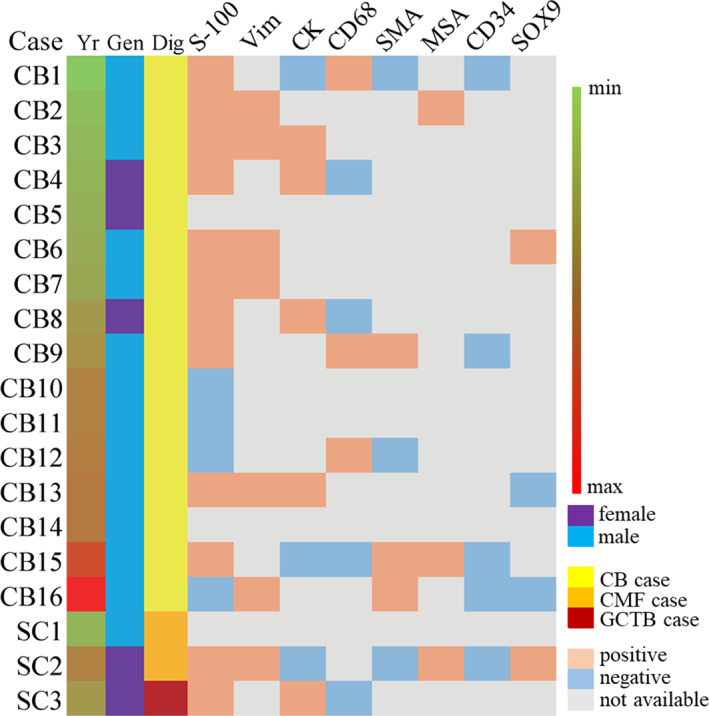
We reviewed and compared our clinical experiences of CB cases and some suspected cases, which were not diagnosed distinctly, between 2013 to 2019. A total of 15 male and four female cases included in this study were seperated into two groups, CB group and suspected case (SC) group. The CB group included 13 men and 3 women, with an age range from 9 to 54 (mean age, 22 years old). The SC group included two men and one woman, with the age range from 13 to 25 (mean age, 19 years old). In both groups the patients had been followed-up until December 2019 and none of the patients had prior treatment history. We evaluated the clinical complaints, radiological features, and clinical-histological features of the cases and performed an immunohistochemical (IHC) study to detect whether the H3K36M expression of cases was different, consistent with a gene-mutation analysis.
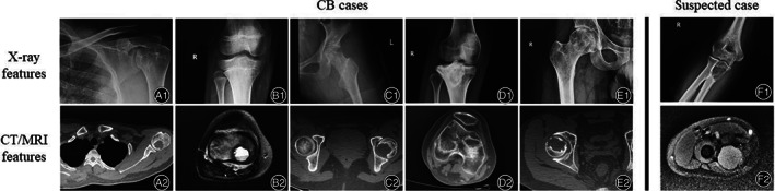
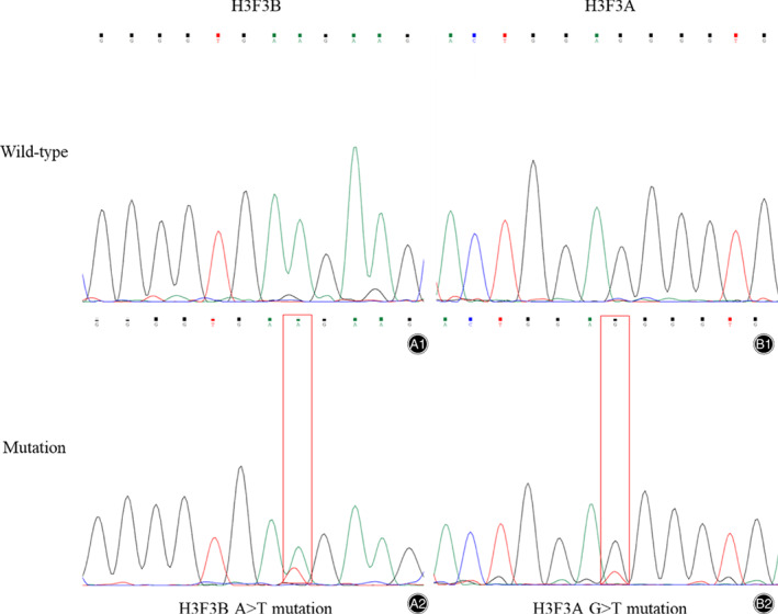
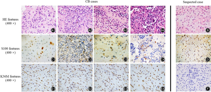
In both groups, the radiologic features of both groups appeared as round low-density shadow with a clear edge, pathologic features showed diffuse proliferation of neoplastic cells with multinuclear giant cells. The radiological tumor size of CB group and SC group showed little difference, which was about 29.0*21.6 mm. Clinical-immunohistochemical features of both groups showed chondroid matrix inside with naïve tumor cells, multinucleated giant cells, and ground substance cells. Most of them showed chondro-related antibody positive (12 cases) but some of them showed S-100 negative (four cases). The clear difference of both groups was the result of H3K36M IHC study and gene analysis. In our cases, the CB group showed diffuse H3K36M positive and the SC group showed negative. The gene mutation analysis revealed that H3K36M-positive CB patients had K36M mutation, which were not found in the SC group. Sanger sequencing showed an A > T substitution at codon 36 of histone H3F3B. No other types of histone H3 mutation was detected in the CB group. Particularly, one of the suspected cases showed a G34W mutation was confirmed to be a giant cell tumor of bone (GCTB).
Our study showed H3K36M immunohistochemistry and gene mutation analysis were specific clinical diagnostic tools to distinguish suspected CB from other giant cell-rich or cartilage matrix-diffuse bone tumors. The clinical-radiological and histomorphological features of patients gave suggestions on whether the H3K36M IHC and gene analysis should be required.
如果软骨母细胞瘤(CB)患者的诊断不明确,且临床上怀疑不明确,H3.3 K36M 突变(H3K36M)能否作为一种方法。
我们回顾性分析了 2013 年至 2019 年间我们的 CB 病例和一些临床怀疑病例的临床经验,这些病例的诊断不明确。本研究共纳入 15 例男性和 4 例女性,分为 CB 组和疑似病例(SC)组。CB 组包括 13 例男性和 3 例女性,年龄 9 至 54 岁(平均年龄 22 岁)。SC 组包括 2 例男性和 1 例女性,年龄 13 至 25 岁(平均年龄 19 岁)。两组患者均随访至 2019 年 12 月,均无既往治疗史。我们评估了病例的临床症状、影像学特征和临床组织学特征,并进行了免疫组织化学(IHC)研究,以检测病例的 H3K36M 表达是否不同,是否与基因突变分析一致。
两组的影像学特征均表现为边缘清晰的圆形低密度影,病理特征均表现为肿瘤细胞弥漫性增生,伴有多核巨细胞。CB 组和 SC 组的肿瘤大小无明显差异,约为 29.0*21.6mm。两组的临床免疫组织化学特征均表现为软骨基质内幼稚肿瘤细胞、多核巨细胞和基质细胞。大多数病例表现为软骨相关抗体阳性(12 例),但部分病例 S-100 阴性(4 例)。两组的明显区别是 H3K36M IHC 研究和基因分析的结果。在我们的病例中,CB 组表现为弥漫性 H3K36M 阳性,而 SC 组表现为阴性。基因突变分析显示 H3K36M 阳性的 CB 患者存在 H3F3B 密码子 36 处的 K36M 突变,而 SC 组则未发现。在 CB 组中未检测到其他类型的组蛋白 H3 突变。特别地,一个疑似病例显示 G34W 突变,被证实为骨巨细胞瘤(GCTB)。
本研究表明 H3K36M 免疫组化和基因突变分析是鉴别疑似 CB 与其他富含巨细胞或软骨基质弥漫性骨肿瘤的特异性临床诊断工具。患者的临床-影像学和组织形态学特征提示是否需要进行 H3K36M IHC 和基因分析。