• 文献检索
  • 文档翻译
  • 深度研究
  • 学术资讯
  • Suppr Zotero 插件Zotero 插件
  • 邀请有礼
  • 套餐&价格
  • 历史记录
应用&插件
Suppr Zotero 插件Zotero 插件浏览器插件Mac 客户端Windows 客户端微信小程序
定价
高级版会员购买积分包购买API积分包
服务
文献检索文档翻译深度研究API 文档MCP 服务
关于我们
关于 Suppr公司介绍联系我们用户协议隐私条款
关注我们

Suppr 超能文献

核心技术专利:CN118964589B侵权必究
粤ICP备2023148730 号-1Suppr @ 2026

文献检索

告别复杂PubMed语法,用中文像聊天一样搜索,搜遍4000万医学文献。AI智能推荐,让科研检索更轻松。

立即免费搜索

文件翻译

保留排版,准确专业,支持PDF/Word/PPT等文件格式,支持 12+语言互译。

免费翻译文档

深度研究

AI帮你快速写综述,25分钟生成高质量综述,智能提取关键信息,辅助科研写作。

立即免费体验

应变超声心动图能否帮助评估危重症 COVID-19 患者的收缩功能?

Could strain echocardiography help to assess systolic function in critically ill COVID-19 patients?

机构信息

Intensive Care Department, Hospital Garcia de Orta, Almada, Portugal.

MiCo, Consulting & Research, Denens, Switzerland.

出版信息

J Clin Monit Comput. 2021 Oct;35(5):1229-1234. doi: 10.1007/s10877-021-00677-1. Epub 2021 Feb 27.

DOI:10.1007/s10877-021-00677-1
PMID:33638061
原文链接:https://pmc.ncbi.nlm.nih.gov/articles/PMC7910195/
Abstract

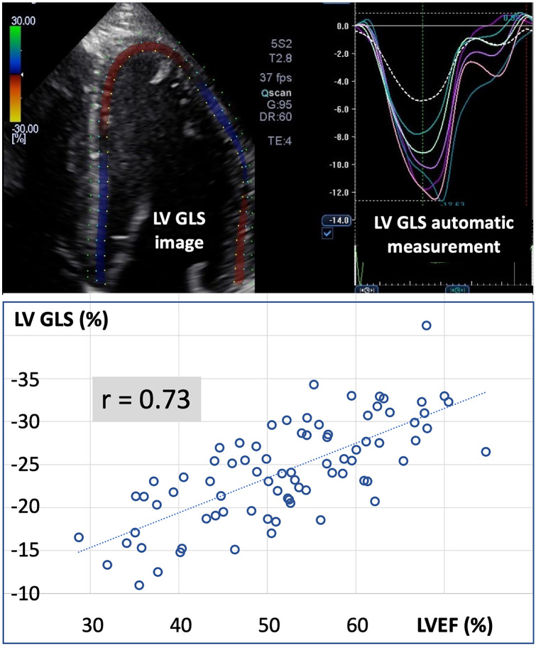
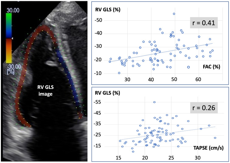
Strain echocardiography enables the automatic quantification of the global longitudinal strain (GLS), which is a direct measure of ventricular shortening during systole. In the current context of overwhelmed ICUs and clinician shortage, GLS has the advantage to be quick and easy to measure by non-experts. However, little is known regarding its value to assess bi-ventricular systolic function in critically ill COVID-19 patients. Therefore, we designed a study to compare right and left ventricular GLS with classic echo-Doppler indices of systolic function, namely the ejection fraction for the left ventricle (LVEF) and the fractional area change (FAC), the tricuspid annular plane systolic excursion (TAPSE), and the tissue Doppler velocity of the basal free lateral wall (S') for the right ventricle. Eighty transthoracic echocardiographic evaluations done in 30 ICU patients with COVID-19 were analyzed. We observed a fair relationship (r = 0.73, p < 0.01) between LVEF and left ventricular GLS. The GLS cut-off value of - 22% identified a LVEF < 50% with a sensitivity of 63% and a specificity of 80%. All patients with a GLS > - 17% had a LVEF < 50%. Although statistically significant, relationships between FAC (r = 0.41, p < 0.01), TAPSE (r = 0.26, p < 0.05) and right ventricular GLS were weak. S' was not correlated with right ventricular GLS. In conclusion, left ventricular GLS was useful to assess left ventricular systolic function. However, right ventricular GLS was poorly correlated with FAC, TAPSE and S'. Further studies are needed to clarify what is the best method to assess right ventricular systolic function in ICU patients with COVID-19.

摘要

应变超声心动图能够自动量化整体纵向应变(GLS),这是心室在收缩期缩短的直接测量指标。在当前 ICU 人满为患和临床医生短缺的情况下,GLS 的优势在于非专业人员可以快速、轻松地测量。然而,对于评估 COVID-19 危重症患者的双心室收缩功能,其价值知之甚少。因此,我们设计了一项研究,比较右心室和左心室 GLS 与经典超声心动图收缩功能指标,即左心室射血分数(LVEF)和左心室的分数面积变化(FAC)、三尖瓣环平面收缩期位移(TAPSE)和右心室基底部游离侧壁的组织多普勒速度(S')。对 30 例 COVID-19 危重症患者的 80 次经胸超声心动图评估进行了分析。我们观察到 LVEF 与左心室 GLS 之间存在良好的相关性(r = 0.73,p < 0.01)。GLS 截断值为-22%时,LVEF<50%的灵敏度为 63%,特异性为 80%。所有 GLS > -17%的患者 LVEF<50%。尽管具有统计学意义,但 FAC(r = 0.41,p < 0.01)和 TAPSE(r = 0.26,p < 0.05)与右心室 GLS 之间的关系较弱。S'与右心室 GLS 不相关。总之,左心室 GLS 有助于评估左心室收缩功能。然而,右心室 GLS 与 FAC、TAPSE 和 S'相关性较差。需要进一步的研究来阐明在 COVID-19 危重症患者中评估右心室收缩功能的最佳方法。

https://cdn.ncbi.nlm.nih.gov/pmc/blobs/1e86/7910195/0fcd4b6889b0/10877_2021_677_Fig2_HTML.jpg
https://cdn.ncbi.nlm.nih.gov/pmc/blobs/1e86/7910195/e5002d605c2c/10877_2021_677_Fig1_HTML.jpg
https://cdn.ncbi.nlm.nih.gov/pmc/blobs/1e86/7910195/0fcd4b6889b0/10877_2021_677_Fig2_HTML.jpg
https://cdn.ncbi.nlm.nih.gov/pmc/blobs/1e86/7910195/e5002d605c2c/10877_2021_677_Fig1_HTML.jpg
https://cdn.ncbi.nlm.nih.gov/pmc/blobs/1e86/7910195/0fcd4b6889b0/10877_2021_677_Fig2_HTML.jpg

相似文献

1
Could strain echocardiography help to assess systolic function in critically ill COVID-19 patients?应变超声心动图能否帮助评估危重症 COVID-19 患者的收缩功能?
J Clin Monit Comput. 2021 Oct;35(5):1229-1234. doi: 10.1007/s10877-021-00677-1. Epub 2021 Feb 27.
2
Echocardiographic predictors of intraoperative right ventricular dysfunction: a 2D and speckle tracking echocardiography study.术中右心室功能障碍的超声心动图预测指标:一项二维和斑点追踪超声心动图研究
Cardiovasc Ultrasound. 2019 Jun 7;17(1):11. doi: 10.1186/s12947-019-0161-3.
3
Usefulness of Right Ventricular Longitudinal Shortening Fraction to Detect Right Ventricular Dysfunction in Acute Cor Pulmonale Related to COVID-19.右心室纵向缩短分数在检测 COVID-19 相关急性肺心病右心功能障碍中的作用。
J Cardiothorac Vasc Anesth. 2021 Dec;35(12):3594-3603. doi: 10.1053/j.jvca.2021.01.025. Epub 2021 Jan 18.
4
Traditional and innovative echocardiographic parameters for the analysis of right ventricular performance in comparison with cardiac magnetic resonance.传统和创新的超声心动图参数用于分析右心室功能,并与心脏磁共振进行比较。
Eur Heart J Cardiovasc Imaging. 2015 Jan;16(1):47-52. doi: 10.1093/ehjci/jeu156. Epub 2014 Sep 3.
5
The evaluation of right ventricular systolic function in patients with repaired Tetralogy of Fallot by conventional echocardiographic methods and speckle tracking echocardiography: Compared with the gold standard cardiac mangenetic resonance.采用传统超声心动图方法和斑点追踪超声心动图评估法洛四联症修复术后患者的右心室收缩功能:与金标准心脏磁共振成像对比
Echocardiography. 2019 Dec;36(12):2251-2258. doi: 10.1111/echo.14532. Epub 2019 Nov 22.
6
Biventricular Function is Impaired in Right Ventricular Septal Pacing-A Prospective Study Using Myocardial Strain Imaging.右室间隔部起搏导致双心室功能障碍:一项应用心肌应变成像的前瞻性研究。
Heart Lung Circ. 2023 Mar;32(3):373-378. doi: 10.1016/j.hlc.2022.11.004. Epub 2022 Dec 20.
7
The value of speckle-tracking echocardiography in identifying right heart dysfunction in patients with chronic thromboembolic pulmonary hypertension.斑点追踪超声心动图在识别慢性血栓栓塞性肺动脉高压患者右心功能不全中的价值。
Int J Cardiovasc Imaging. 2018 Dec;34(12):1895-1904. doi: 10.1007/s10554-018-1423-0. Epub 2018 Jul 30.
8
Tricuspid annular plane systolic excursion and central venous pressure in mechanically ventilated critically ill patients.机械通气的危重症患者的三尖瓣环平面收缩期位移与中心静脉压
Cardiovasc Ultrasound. 2018 Aug 7;16(1):11. doi: 10.1186/s12947-018-0130-2.
9
Serial Changes in Right Ventricular Systolic Function Among Rejection-Free Children and Young Adults After Heart Transplantation.心脏移植后排斥反应阴性的儿童和青年右心室收缩功能的连续变化。
J Am Soc Echocardiogr. 2019 Aug;32(8):1027-1035.e2. doi: 10.1016/j.echo.2019.04.413. Epub 2019 Jun 13.
10
Assessment of right ventricular systolic function in heart failure with preserved, reduced and mid-range ejection fraction.射血分数保留、降低和中等范围的心力衰竭患者右心室收缩功能评估
Indian Heart J. 2019 Sep-Oct;71(5):406-411. doi: 10.1016/j.ihj.2019.11.252. Epub 2019 Nov 20.

引用本文的文献

1
AI for the hemodynamic assessment of critically ill and surgical patients: focus on clinical applications.人工智能在危重症及外科手术患者血流动力学评估中的应用:聚焦临床应用
Ann Intensive Care. 2025 Feb 24;15(1):26. doi: 10.1186/s13613-025-01448-w.
2
Visualizing hemodynamics: innovative graphical displays and imaging techniques in anesthesia and critical care.可视化血流动力学:麻醉与重症监护中的创新图形显示与成像技术
Crit Care. 2025 Jan 3;29(1):3. doi: 10.1186/s13054-024-05239-w.
3
Past and Present of Point-of-Care Ultrasound (PoCUS): A Narrative Review.

本文引用的文献

1
Echocardiographic features of patients with COVID-19 infection: a cross-sectional study.COVID-19 感染患者的超声心动图特征:一项横断面研究。
Int J Cardiovasc Imaging. 2021 Mar;37(3):825-834. doi: 10.1007/s10554-020-02051-9. Epub 2020 Oct 8.
2
Critically ill patients with COVID-19: are they hemodynamically unstable and do we know why?新型冠状病毒肺炎危重症患者:他们血流动力学不稳定吗?我们知道原因吗?
Intensive Care Med. 2021 Feb;47(2):254-255. doi: 10.1007/s00134-020-06238-5. Epub 2020 Sep 10.
3
Perioperative echocardiography-guided hemodynamic therapy in high-risk patients: a practical expert approach of hemodynamically focused echocardiography.
床旁超声(PoCUS)的过去与现在:一篇叙述性综述
Cureus. 2023 Dec 8;15(12):e50155. doi: 10.7759/cureus.50155. eCollection 2023 Dec.
4
Echocardiography in Coronavirus Disease 2019 Era: A Single Tool for Diagnosis and Prognosis.2019年冠状病毒病时代的超声心动图:诊断与预后的单一工具
J Cardiovasc Echogr. 2023 Jan-Mar;33(1):10-16. doi: 10.4103/jcecho.jcecho_11_23. Epub 2023 May 29.
5
Transthoracic echocardiography of patients in prone position ventilation during the COVID-19 pandemic: an observational and retrospective study.COVID-19 大流行期间俯卧位通气患者的经胸超声心动图:一项观察性和回顾性研究。
Int J Cardiovasc Imaging. 2022 Nov;38(11):2303-2309. doi: 10.1007/s10554-022-02659-z. Epub 2022 Jun 26.
6
Abnormal Echocardiographic Findings in Hospitalized Patients with Covid-19: A Systematic Review and Meta-analysis.住院新冠患者的超声心动图异常表现:系统评价和荟萃分析。
Arq Bras Cardiol. 2022 Aug;119(2):267-279. doi: 10.36660/abc.20210485.
7
Characteristic Immune Dynamics in COVID-19 Patients with Cardiac Dysfunction.新冠病毒感染合并心脏功能障碍患者的特征性免疫动力学
J Clin Med. 2022 Mar 28;11(7):1880. doi: 10.3390/jcm11071880.
8
The Right Ventricle in COVID-19.新型冠状病毒肺炎中的右心室
J Clin Med. 2021 Jun 8;10(12):2535. doi: 10.3390/jcm10122535.
围术期超声心动图引导高危患者血流动力学治疗:血流动力学聚焦超声心动图的实用专家方法。
J Clin Monit Comput. 2021 Apr;35(2):229-243. doi: 10.1007/s10877-020-00534-7. Epub 2020 May 26.
4
Prognostic Value of Right Ventricular Dysfunction in Heart Failure With Reduced Ejection Fraction: Superiority of Longitudinal Strain Over Tricuspid Annular Plane Systolic Excursion.射血分数降低型心力衰竭患者右心室功能障碍的预后价值:纵向应变优于三尖瓣环平面收缩期位移。
Circ Cardiovasc Imaging. 2018 Jan;11(1):e006894. doi: 10.1161/CIRCIMAGING.117.006894.