Department of Pediatrics, Division of Pediatric Cardiology, University of Tennessee Health Science Center, Le Bonheur Children's Hospital, 49N Dunlap Street, 3rd Floor, Memphis, TN, 38015, USA.
Department of Pediatrics, Division of Pediatric Cardiology, University of Texas Southwestern Medical Center, Dallas Children's Medical Center, Dallas, TX, USA.
J Cardiovasc Magn Reson. 2021 Mar 1;23(1):16. doi: 10.1186/s12968-021-00707-6.
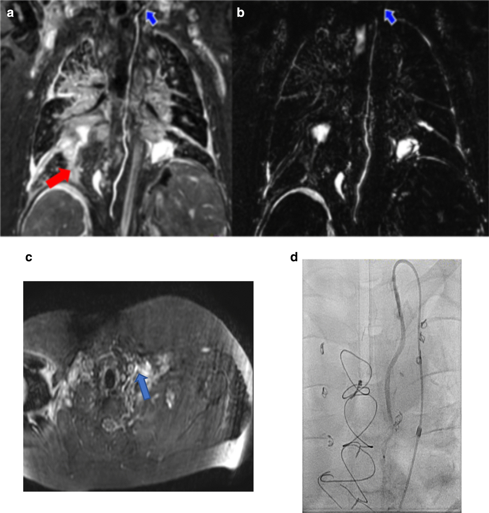
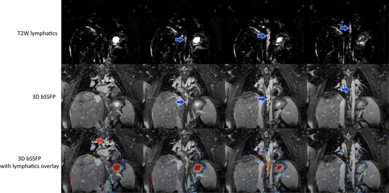
Due to passive blood flow in palliated single ventricle, central venous pressure increases chronically, ultimately impeding lymphatic drainage. Early visualization and treatment of these malformations is essential to reduce morbidity and mortality. Cardiovascular magnetic resonance (CMR) T2-weighted lymphangiography (T2w) is used for lymphatic assessment, but its low signal-to-noise ratio may result in incomplete visualization of thoracic duct pathway. 3D-balanced steady state free precession (3D-bSSFP) is commonly used to assess congenital cardiac disease anatomy. Here, we aimed to improve diagnostic imaging of thoracic duct pathway using 3D-bSSFP.
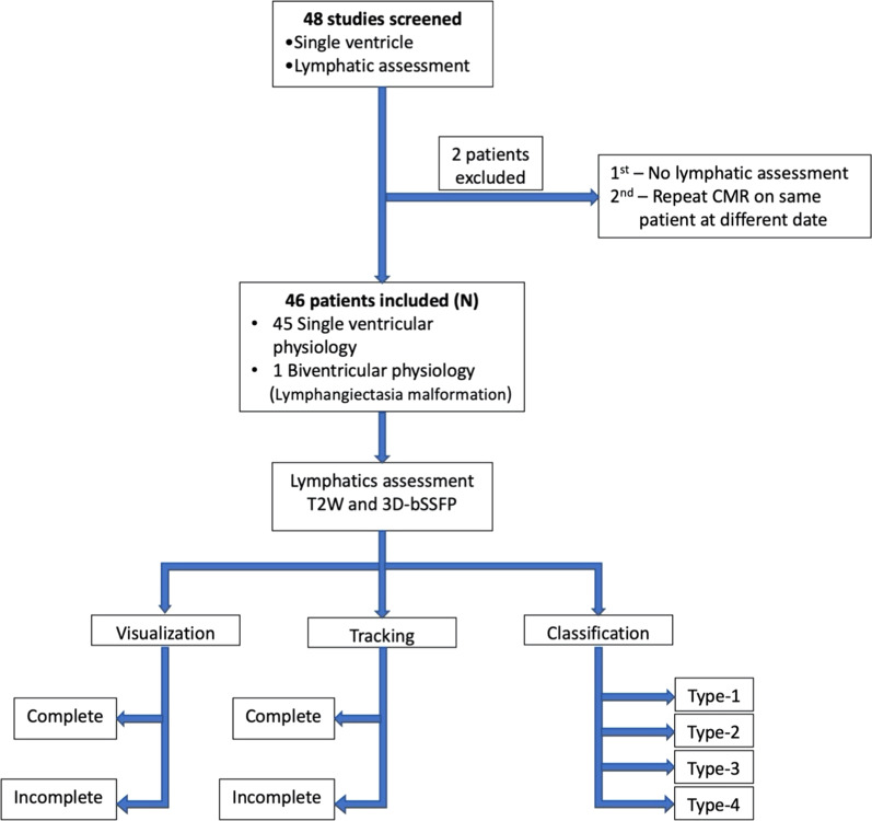
Patients underwent CMR during single ventricle or central lymphatic system assessment using T2w and 3D-bSSFP. T2w parameters included 3D-turbo spin echo (TSE), TE/TR = 600/2500 ms, resolution = 1 × 1 × 1.8 mm, respiratory triggering with bellows. 3D-bSSFP parameters included electrocardiogram triggering and diaphragm navigator, 1.6 mm isotropic resolution, TE/TR = 1.8/3.6 ms. Thoracic duct was identified independently in T2w and 3D-bSSFP images, tracked completely from cisterna chyli to its drainage site, and classified based on severity of lymphatic abnormalities.
Forty-eight patients underwent CMR, 46 of whom were included in the study. Forty-five had congenital heart disease with single ventricle physiology. Median age at CMR was 4.3 year (range 0.9-35.1 year, IQR 2.4 year), and median weight was 14.4 kg (range, 7.9-112.9 kg, IQR 5.2 kg). Single ventricle with right dominant ventricle was noted in 31 patients. Thirty-eight patients (84%) were status post bidirectional Glenn and 7 (16%) were status post Fontan anastomosis. Thoracic duct visualization was achieved in 45 patients by T2w and 3D-bSSFP. Complete tracking to drainage site was attained in 11 patients (24%) by T2w vs 25 (54%) by 3D-bSSFP and in 28 (61%) by both. Classification of lymphatics was performed in 31 patients.
Thoracic duct pathway can be visualized by 3D-bSSFP combined with T2w lymphangiography. Cardiac triggering and respiratory navigation likely help retain lymphatic signal in the retrocardiac area by 3D-bSSFP. Visualizing lymphatic system leaks is challenging on 3D-bSSFP images alone, but 3D-bSSFP offers good visualization of duct anatomy and landmark structures to help plan interventions. Together, these sequences can define abnormal lymphatic pathway following single ventricle palliative surgery, thus guiding lymphatic interventional procedures.
由于姑息性单心室中的被动血流,中心静脉压会长期升高,最终阻碍淋巴引流。早期发现和治疗这些畸形对于降低发病率和死亡率至关重要。心血管磁共振(CMR)T2 加权淋巴管造影术(T2w)用于评估淋巴管,但它的信噪比低,可能导致胸导管途径的不完全可视化。3D 平衡稳态自由进动(3D-bSSFP)常用于评估先天性心脏病解剖结构。在这里,我们旨在使用 3D-bSSFP 改善胸导管途径的诊断成像。
患者在单心室或中央淋巴系统评估期间接受 CMR,使用 T2w 和 3D-bSSFP。T2w 参数包括 3D 涡轮自旋回波(TSE),TE/TR=600/2500ms,分辨率=1×1×1.8mm,呼吸触发用风箱。3D-bSSFP 参数包括心电图触发和膈肌导航,1.6mm 各向同性分辨率,TE/TR=1.8/3.6ms。在 T2w 和 3D-bSSFP 图像中独立识别胸导管,从 cisterna chyli 完全追踪到其引流部位,并根据淋巴管异常的严重程度进行分类。
48 名患者接受了 CMR,其中 46 名患者纳入了研究。45 名患者患有先天性心脏病,单心室生理。CMR 的中位年龄为 4.3 岁(范围 0.9-35.1 岁,IQR 2.4 岁),中位体重为 14.4kg(范围 7.9-112.9kg,IQR 5.2kg)。31 名患者为右优势型右心室。38 名患者(84%)接受了双向 Glenn 手术,7 名患者(16%)接受了 Fontan 吻合术。T2w 和 3D-bSSFP 均可在 45 名患者中实现胸导管可视化。11 名患者(24%)通过 T2w 可完全追踪至引流部位,25 名患者(54%)通过 3D-bSSFP 可完全追踪至引流部位,28 名患者(61%)通过两者均可完全追踪至引流部位。在 31 名患者中进行了淋巴管分类。
3D-bSSFP 结合 T2w 淋巴管造影术可显示胸导管途径。心脏触发和呼吸导航可能有助于通过 3D-bSSFP 在心脏后区保留淋巴管信号。单独使用 3D-bSSFP 很难观察到淋巴管漏,但 3D-bSSFP 可很好地显示导管解剖结构和地标结构,有助于计划干预措施。这些序列可用于定义单心室姑息手术后异常的淋巴途径,从而指导淋巴介入治疗。