Yang Di, Shen Lin-Xia, Chen Ri-Feng, Fu Yu, Xu Hong-Yan, Zhang Li-Na, Liu Dong-Hua
Department of Dermatology, The First Affiliated Hospital of Guangxi Medical University, Nanning, 530021, People's Republic of China.
Department of Dermatology and Venereology, Huashan Hospital, Fudan University, Shanghai, 200040, People's Republic of China.
Infect Drug Resist. 2021 Feb 19;14:651-660. doi: 10.2147/IDR.S297160. eCollection 2021.
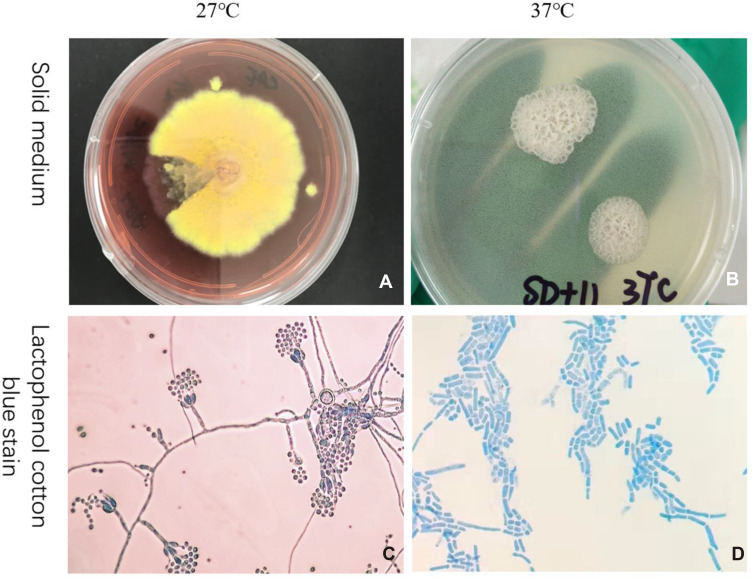
() is a destructive opportunistic dimorphic fungal which can cause lethiferous Talaromycosis, but the clearance of mainly depends on the innate immune response.
To investigate whether can inhibit the expression of CD86 in THP-1 cells after infection and discuss the potential mechanisms.
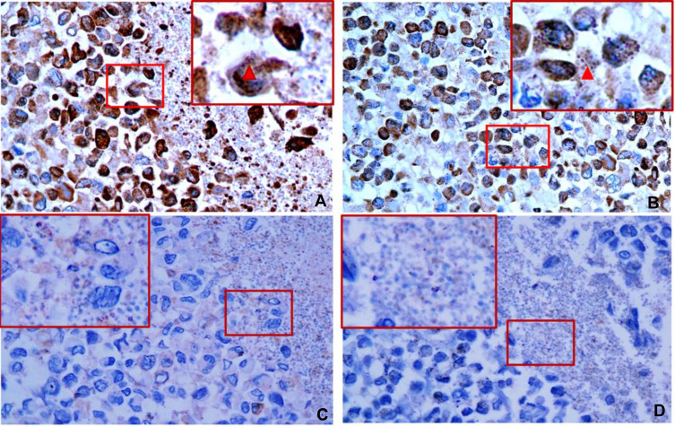
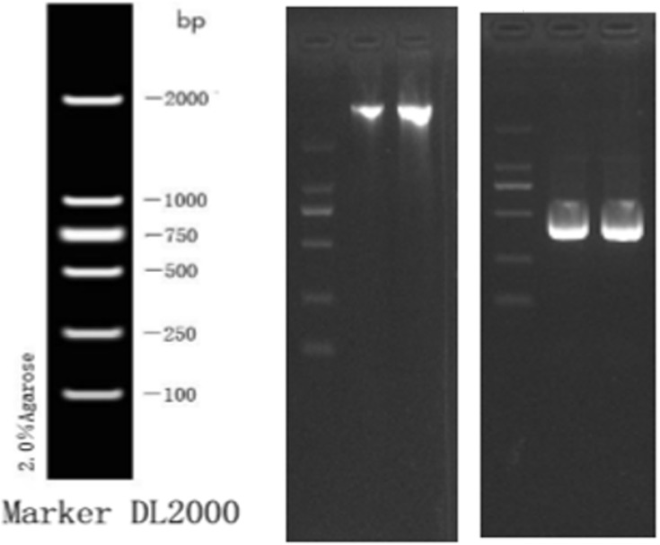
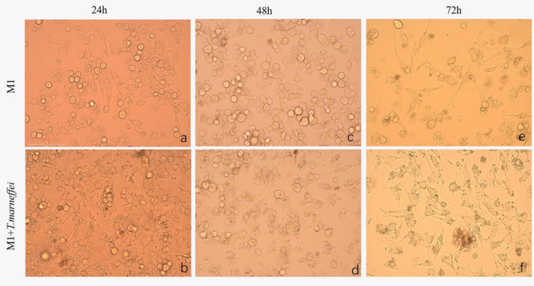
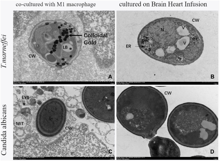
Western blot and immunoelectron microscopy were used to detect the CD86 expression on cultured on BHI medium at 37°C. Western blot, enzyme-linked immunoassay and immunofluorescence were used to detect the change of CD86 expression on macrophages incubating with . Enzyme-linked immunoassay was used to detect the content of CD86 in supernatant in the co-culture system. Immunohistochemistry and immunoelectron microscopy were used to detect the expression of CD86 on incubating with macrophages.
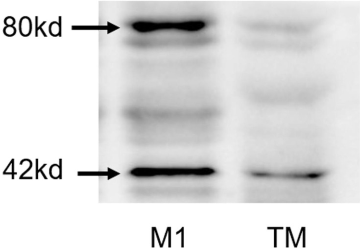
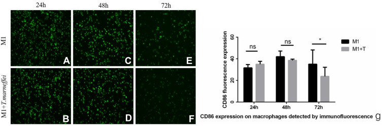
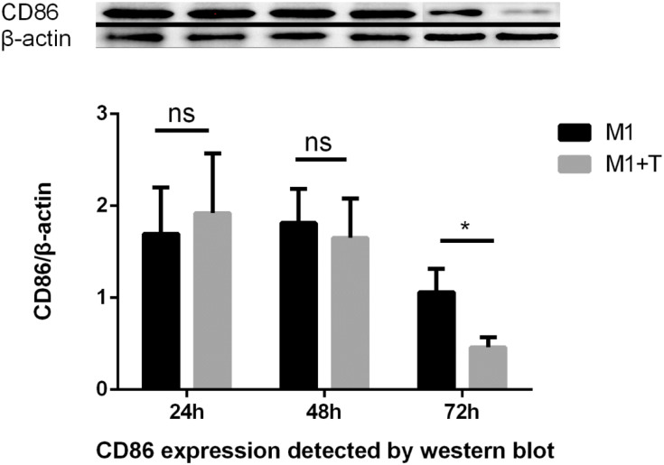
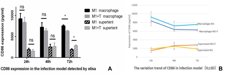
did not express CD86 when cultured separately at 37°C detected by Western blot and immunoelectron microscopy, but it did express CD86 when incubated with macrophages detected by immunohistochemistry and immunoelectron microscopy. The CD86 expression of macrophages significantly decreased at 72 hours when infected with while the content of CD86 in supernatant significantly increased at 72 hours compared with the control group which were detected by Western blot, enzyme-linked immunoassay and immunofluorescence.
()是一种具有破坏性的机会性双相真菌,可引起致死性的嗜热栖热放线菌病,但(该菌的清除)主要依赖于固有免疫反应。
探讨(该菌)感染后是否能抑制THP - 1细胞中CD86的表达,并探讨其潜在机制。
采用蛋白质免疫印迹法和免疫电子显微镜检测在37℃BHI培养基上培养的(该菌)上CD86的表达。采用蛋白质免疫印迹法、酶联免疫吸附测定法和免疫荧光法检测与(该菌)共孵育的巨噬细胞上CD86表达的变化。采用酶联免疫吸附测定法检测共培养体系上清液中CD86的含量。采用免疫组织化学和免疫电子显微镜检测与巨噬细胞共孵育的(该菌)上CD86的表达。
通过蛋白质免疫印迹法和免疫电子显微镜检测,在37℃单独培养时(该菌)不表达CD86,但通过免疫组织化学和免疫电子显微镜检测,与巨噬细胞共孵育时(该菌)表达CD86。蛋白质免疫印迹法、酶联免疫吸附测定法和免疫荧光法检测显示,感染(该菌)后72小时,巨噬细胞的CD86表达显著降低,而与对照组相比,上清液中CD86的含量在72小时显著增加。
1)(该菌)感染后,THP - 1上的CD86表达降低,且随着感染的进展,M1巨噬细胞极化不足逐渐显现;2)(该菌)在与THP - 1细胞接触过程中可能吸附或摄取巨噬细胞产生的上清液中的CD86,从而导致巨噬细胞中CD86的消耗。
原文括号处内容缺失,译文按原文格式保留括号。