Danyluk Hayden, Ishaque Abdullah, Ta Daniel, Yang Yee Hong, Wheatley B Matthew, Kalra Sanjay, Sankar Tejas
Division of Surgical Research, Department of Surgery, University of Alberta, Edmonton, AB, Canada.
Division of Neurosurgery, Department of Surgery, University of Alberta Hospital, University of Alberta, Edmonton, AB, Canada.
Front Neurol. 2021 Feb 12;12:626504. doi: 10.3389/fneur.2021.626504. eCollection 2021.
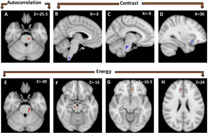
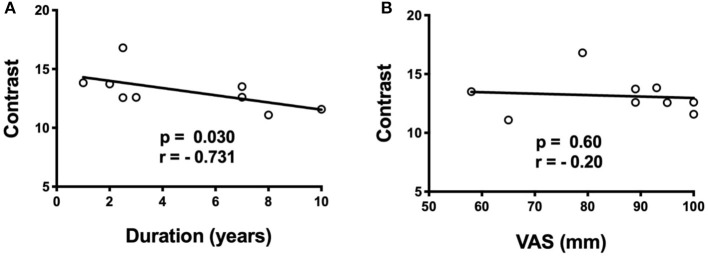
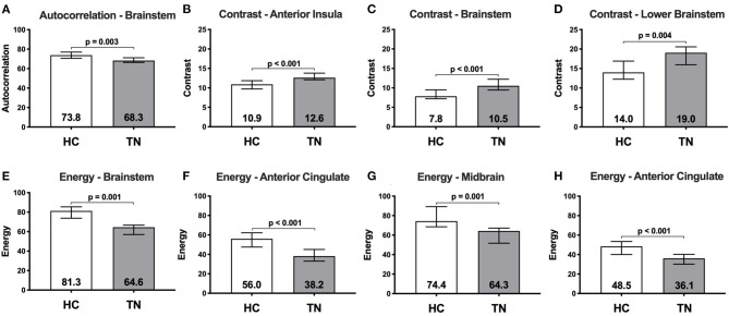
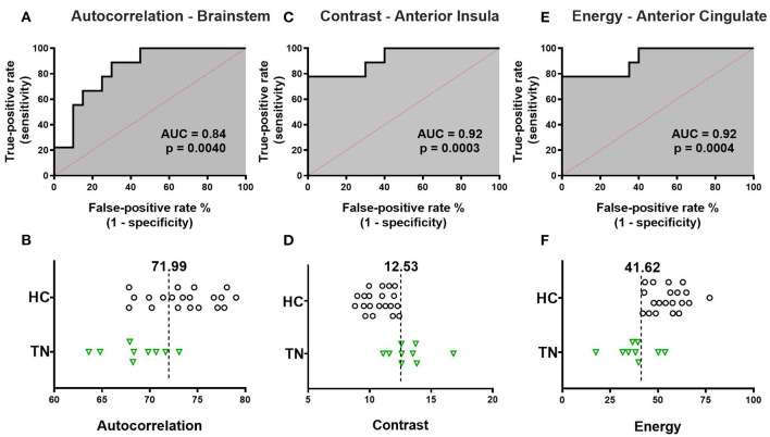
Several neuroimaging studies report structural alterations of the trigeminal nerve in trigeminal neuralgia (TN). Less attention has been paid to structural brain changes occurring in TN, even though such changes can influence the development and response to treatment of other headache and chronic pain conditions. The purpose of this study was to apply a novel neuroimaging technique-texture analysis-to identify structural brain differences between classical TN patients and healthy subjects. We prospectively recruited 14 medically refractory classical TN patients and 20 healthy subjects. 3-Tesla T1-weighted brain MRI scans were acquired in all participants. Three texture features (autocorrelation, contrast, energy) were calculated within four brain regions of interest (anterior cingulate, insula, thalamus, brainstem). Voxel-wise analysis was used to identify clusters of texture difference between TN patients and healthy subjects within regions of interest ( < 0.001, cluster size >20 voxels). Median raw texture values within clusters were also compared between groups, and further used to differentiate TN patients from healthy subjects (receiver-operator characteristic curve analysis). Median raw texture values were correlated with pain severity (visual analog scale, 1-100) and illness duration. Several clusters of texture difference were observed between TN patients and healthy subjects: right-sided TN patients showed reduced autocorrelation in the left brainstem, increased contrast in the left brainstem and right anterior insula, and reduced energy in right and left anterior cingulate, right midbrain, and left brainstem. Within-cluster median raw texture values also differed between TN patients and healthy subjects: TN patients could be segregated from healthy subjects using brainstem autocorrelation ( = 0.0040, AUC = 0.84, sensitivity = 89%, specificity = 70%), anterior insula contrast ( = 0.0002, AUC = 0.92, sensitivity = 78%, specificity = 100%), and anterior cingulate energy ( = 0.0004, AUC = 0.92, sensitivity = 78%, specificity = 100%). Additionally, anterior insula contrast and duration of TN were inversely correlated ( = 0.030, Spearman r = -0.73). Texture analysis reveals distinct brain abnormalities in TN, which relate to clinical features such as duration of illness. These findings further implicate structural brain changes in the development and maintenance of TN.
多项神经影像学研究报告了三叉神经痛(TN)患者三叉神经的结构改变。尽管这些改变会影响其他头痛和慢性疼痛疾病的发展及治疗反应,但TN患者脑部的结构变化却较少受到关注。本研究旨在应用一种新型神经影像学技术——纹理分析,来识别典型TN患者与健康受试者之间的脑部结构差异。我们前瞻性招募了14例药物难治性典型TN患者和20名健康受试者。所有参与者均接受了3特斯拉T1加权脑部MRI扫描。在四个感兴趣的脑区(前扣带回、岛叶、丘脑、脑干)计算了三种纹理特征(自相关、对比度、能量)。采用体素分析来识别TN患者与健康受试者在感兴趣区域内的纹理差异簇(<0.001,簇大小>20体素)。还比较了两组之间簇内的原始纹理值中位数,并进一步用于区分TN患者与健康受试者(受试者工作特征曲线分析)。原始纹理值中位数与疼痛严重程度(视觉模拟量表,1 - 100)和病程相关。在TN患者与健康受试者之间观察到了几个纹理差异簇:右侧TN患者左侧脑干的自相关降低,左侧脑干和右侧前岛叶的对比度增加,右侧和左侧前扣带回、右侧中脑和左侧脑干的能量降低。TN患者与健康受试者之间簇内的原始纹理值中位数也存在差异:使用脑干自相关(=0.0040,AUC = 0.84,敏感性 = 89%,特异性 = 70%)、前岛叶对比度(=0.0002,AUC = 0.92,敏感性 = 78%,特异性 = 100%)和前扣带回能量(=0.0004,AUC = 0.92,敏感性 = 78%,特异性 = 100%)可将TN患者与健康受试者区分开来。此外,前岛叶对比度与TN病程呈负相关(=0.030,Spearman r = -0.73)。纹理分析揭示了TN患者脑部存在明显异常,这些异常与病程等临床特征相关。这些发现进一步表明脑部结构变化在TN的发生和维持中起作用。