Xue Jing-Shi, Yao Chi, Xu Qin-Lin, Sui Chang-Xu, Jia Xin-Lei, Hu Wen-Jing, Lv Yong-Lin, Feng Yi-Feng, Peng Yu-Jia, Shen Shi-Yi, Yang Nai-Ying, Lou Yu-Xia, Yang Zhong-Nan
Shanghai Key Laboratory of Plant Molecular Sciences, College of Life Sciences, Shanghai Normal University, Shanghai, China.
Front Plant Sci. 2021 Feb 10;12:634114. doi: 10.3389/fpls.2021.634114. eCollection 2021.
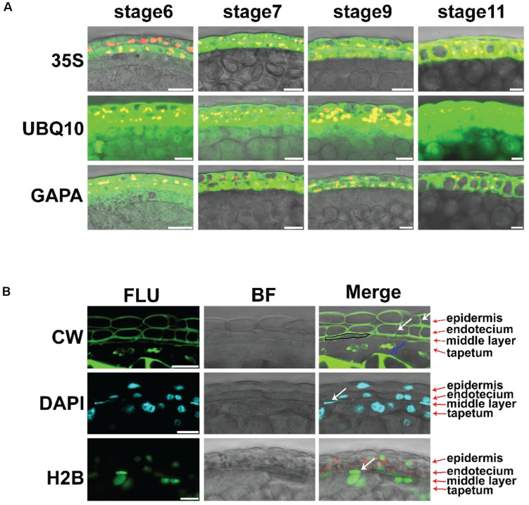
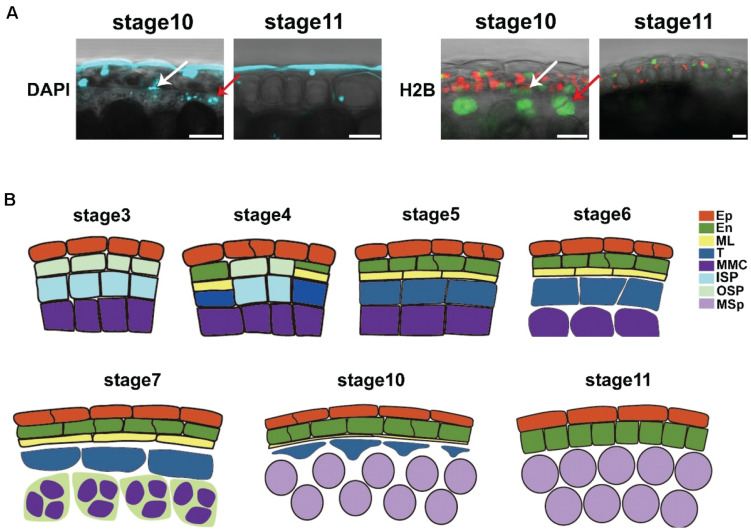
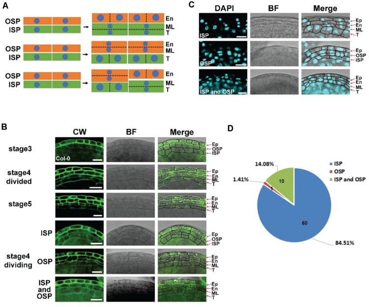
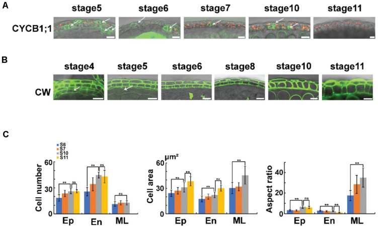
The middle layer is an essential cell layer of the anther wall located between the endothecium and tapetum in . Based on sectioning, the middle layer was found to be degraded at stage 7, which led to the separation of the tapetum from the anther wall. Here, we established techniques for live imaging of the anther. We created a marker line with fluorescent proteins expressed in all anther layers to study anther development. Several staining methods were used in the intact anthers to study anther cell morphology. We clarified the initiation, development, and degradation of the middle layer in . This layer is initiated from both the inner and outer secondary parietal cells at stage 4, stopped cell division at stage 6, and finally degraded at stage 11. The neighboring cell layers, the epidermis, and endothecium continued cell division until stage 10, which led to a thin middle layer. The degradation of the tapetum cell wall at stage 7 lead to its isolation from the anther wall. This work presents fundamental information on the development of the middle layer, which facilitates the further investigation of anther development and plant fertility. These live imaging methods could be useful in future studies.
中间层是花药壁的一个重要细胞层,位于药室内壁和绒毡层之间。通过切片观察发现,中间层在第7阶段开始退化,导致绒毡层与花药壁分离。在此,我们建立了花药活体成像技术。我们创建了一个在所有花药层中表达荧光蛋白的标记系,以研究花药发育。在完整的花药中使用了几种染色方法来研究花药细胞形态。我们阐明了中间层在[具体植物名称未给出]中的起始、发育和退化过程。该层在第4阶段由内、外次生周缘细胞起始,在第6阶段停止细胞分裂,最终在第11阶段退化。相邻的细胞层,即表皮和药室内壁,持续细胞分裂直至第10阶段,这导致中间层变薄。第7阶段绒毡层细胞壁的退化导致其与花药壁分离。这项工作提供了关于中间层发育的基本信息,有助于进一步研究花药发育和植物育性。这些活体成像方法在未来的研究中可能会有用。