Division of Oncology and Christian Doppler Laboratory for Personalized Immunotherapy, Department of Medicine I, Medical University of Vienna, Waehringer Guertel 18-20, 1090, Vienna, Austria.
Department of Neurosurgery, Medical University of Vienna, Vienna, Austria.
J Neurooncol. 2021 May;152(3):533-539. doi: 10.1007/s11060-021-03721-x. Epub 2021 Mar 2.
Immune modulatory therapies including immune checkpoint inhibitors have so far failed to result in clinically meaningful efficacy in glioma. We aimed to investigate lymphocyte activation gene 3 (LAG-3), an inhibitory receptor on immune cells and target of second-generation immune checkpoint inhibitors, in glioma.
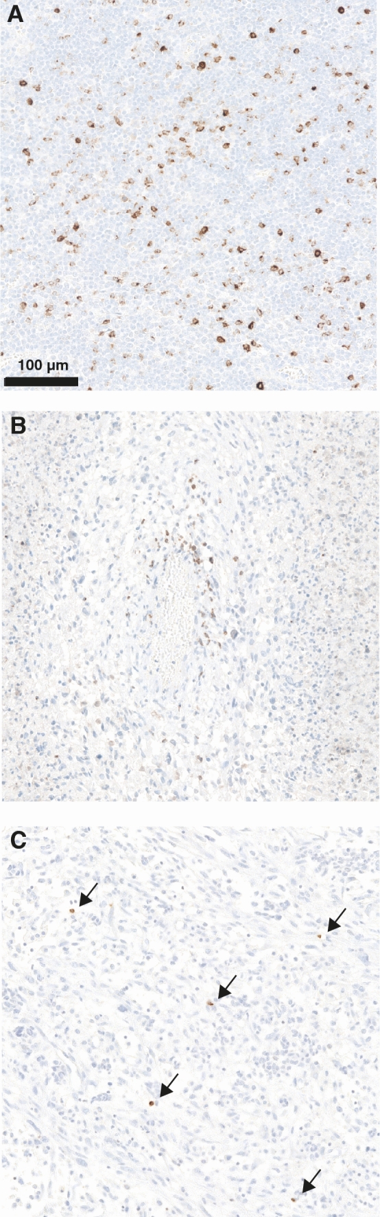
97 patients with diffuse glioma (68 with glioblastoma, 29 with WHO grade II-III glioma) were identified from the Neuro-Biobank of the Medical University of Vienna. LAG-3 expression in the inflammatory microenvironment was assessed by immunohistochemistry (monoclonal anti-LAG-3 antibody, clone 17B4) and correlated to CD3+ , CD8+ , CD20+ and PD-1+ tumor-infiltrating lymphocytes (TILs) and PD-L1 expression on tumor cells.
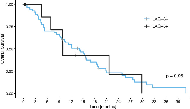
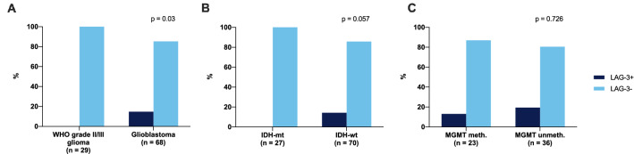
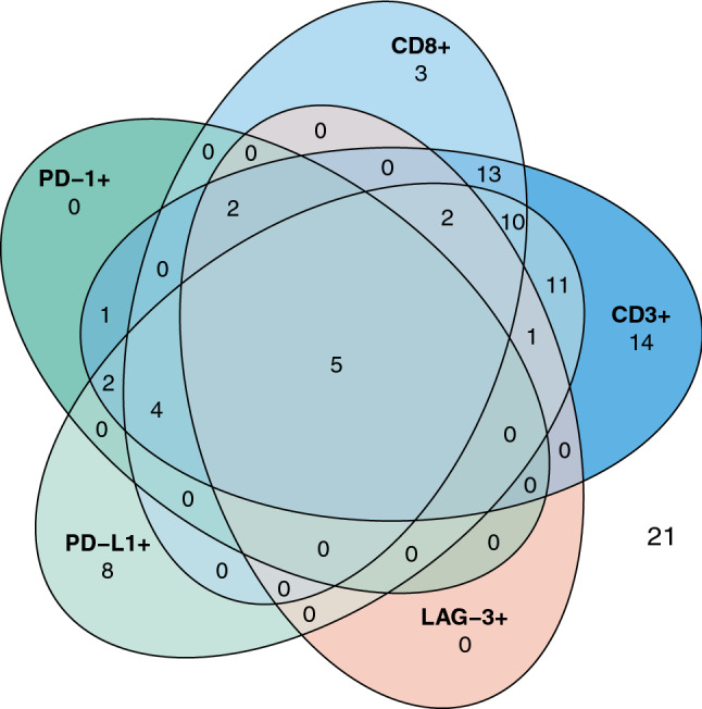
LAG-3+ TILs could be observed in 10/97 (10.3%) IDH-wildtype samples and in none of the included IDH-mutant glioma samples (p = 0.057). Further, LAG-3+ TILs were only observed in WHO grade IV glioblastoma, while none of the investigated WHO grade II-III glioma presented with LAG-3+ TILs (p = 0.03). No association of O6-methylguanine-DNA-methyltransferase (MGMT) promoter methylation and presence of LAG-3+ TILs was observed (p = 0.726). LAG-3 expression was associated with the presence of CD3+ (p = 0.029), CD8+ (p = 0.001), PD-1+ (p < 0.001) TILs and PD-L1+ tumor cells (p = 0.021), respectively. No association of overall survival with LAG-3+ TIL infiltration was evident (median OS 9.9 vs. 14.2 months, p = 0.95).
LAG-3 is only rarely expressed on TILs in IDH-wildtype glioma and associated with active inflammatory milieu as defined by higher TIL density. Immune microenvironment diversity should be considered in the design of future immunotherapy trials in glioma.
免疫调节疗法,包括免疫检查点抑制剂,迄今为止并未在神经胶质瘤中产生有临床意义的疗效。我们旨在研究淋巴细胞激活基因 3(LAG-3),一种免疫细胞上的抑制受体,也是第二代免疫检查点抑制剂的靶点,在神经胶质瘤中的作用。
从维也纳医科大学神经生物库中确定了 97 名弥漫性神经胶质瘤患者(68 名胶质母细胞瘤,29 名 WHO 分级 II-III 级神经胶质瘤)。通过免疫组织化学(单克隆抗 LAG-3 抗体,克隆 17B4)评估炎症微环境中的 LAG-3 表达,并与 CD3+、CD8+、CD20+和 PD-1+肿瘤浸润淋巴细胞(TILs)以及肿瘤细胞上的 PD-L1 表达相关联。
在 10/97(10.3%)IDH 野生型样本中观察到 LAG-3+TILs,而在纳入的 IDH 突变型神经胶质瘤样本中均未观察到(p=0.057)。此外,LAG-3+TILs 仅在 WHO 分级 IV 级胶质母细胞瘤中观察到,而在所研究的 WHO 分级 II-III 级神经胶质瘤中均未观察到 LAG-3+TILs(p=0.03)。O6-甲基鸟嘌呤-DNA-甲基转移酶(MGMT)启动子甲基化与 LAG-3+TILs 的存在之间无相关性(p=0.726)。LAG-3 表达与 CD3+(p=0.029)、CD8+(p=0.001)、PD-1+(p<0.001)TILs 和 PD-L1+肿瘤细胞(p=0.021)的存在相关。LAG-3+TIL 浸润与总生存期无明显相关性(中位 OS 9.9 与 14.2 个月,p=0.95)。
LAG-3 在 IDH 野生型神经胶质瘤的 TIL 中很少表达,与更高的 TIL 密度所定义的活跃炎症环境相关。在神经胶质瘤的未来免疫治疗试验设计中应考虑免疫微环境的多样性。