Skirball Institute of Biomolecular Medicine, New York University Langone Medical Center, New York, NY, USA.
Graduate School of Arts and Sciences, Harvard University, Cambridge, MA, USA.
Nature. 2021 Apr;592(7853):290-295. doi: 10.1038/s41586-021-03227-6. Epub 2021 Mar 3.
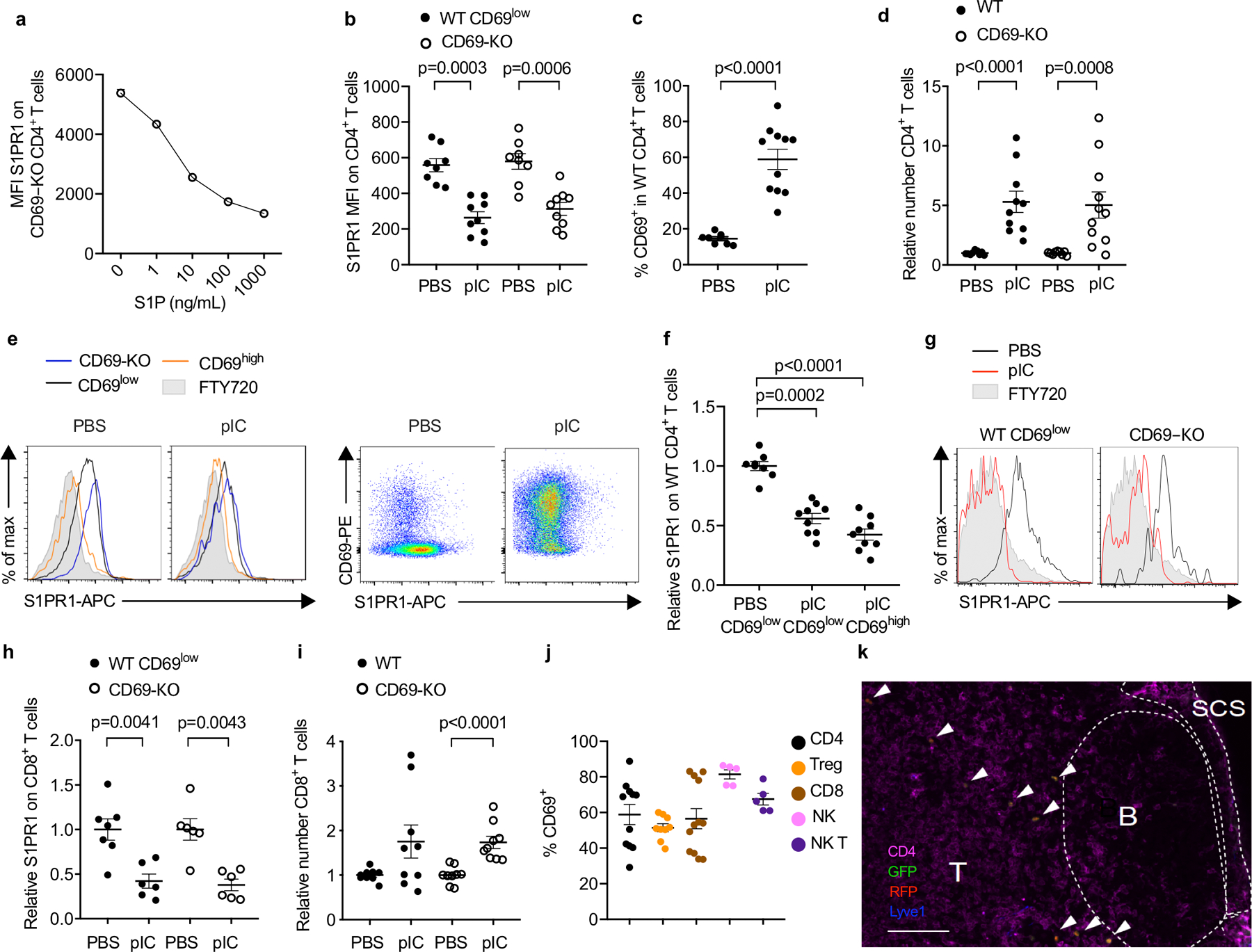
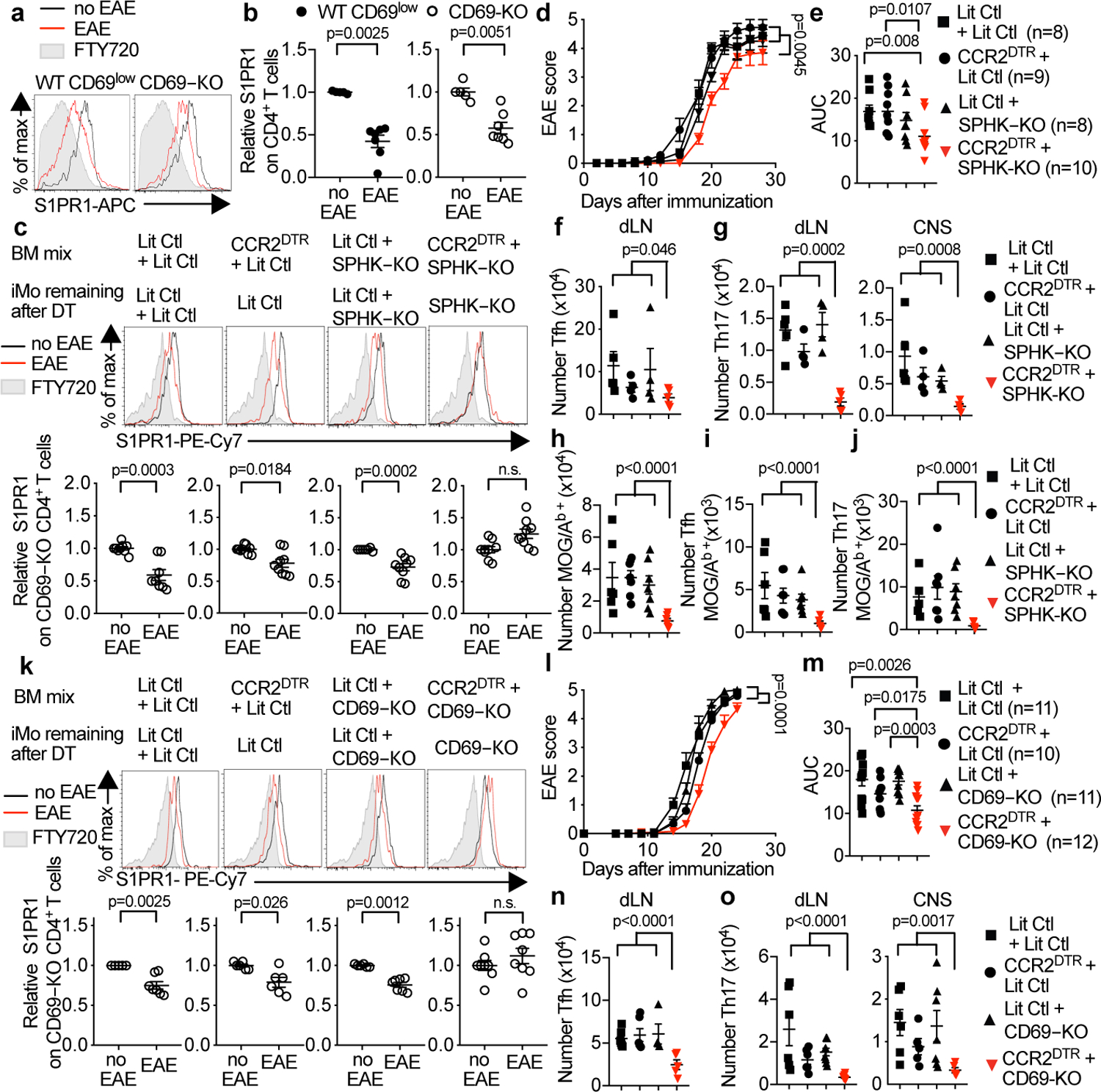
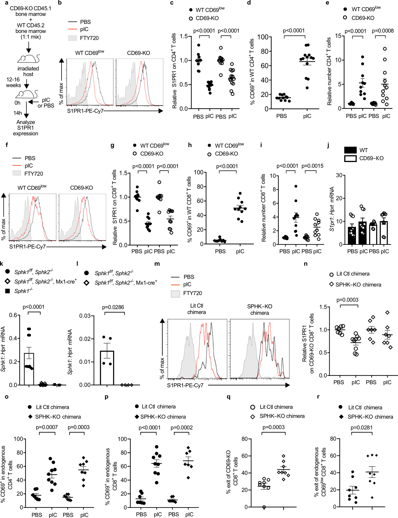
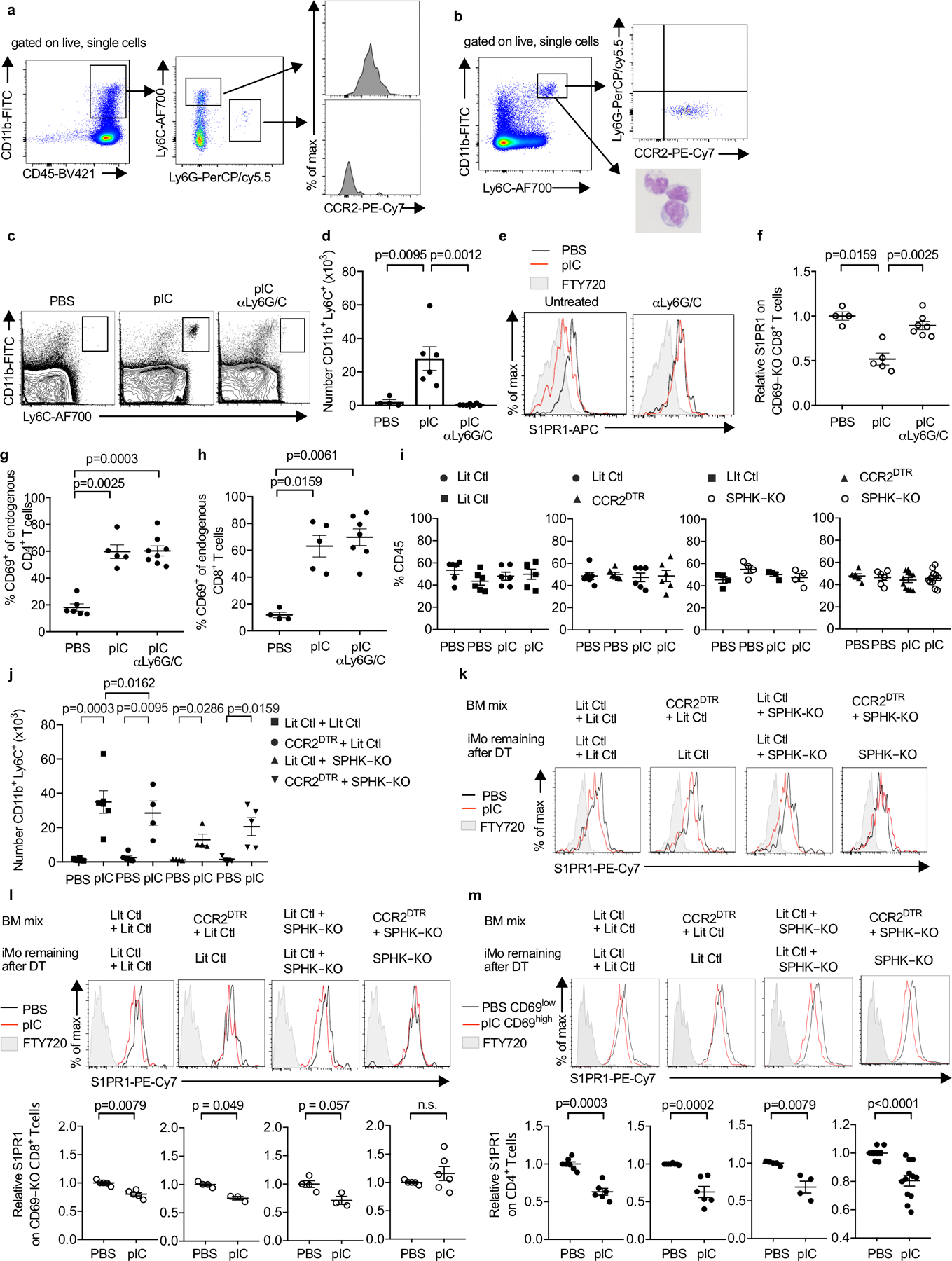
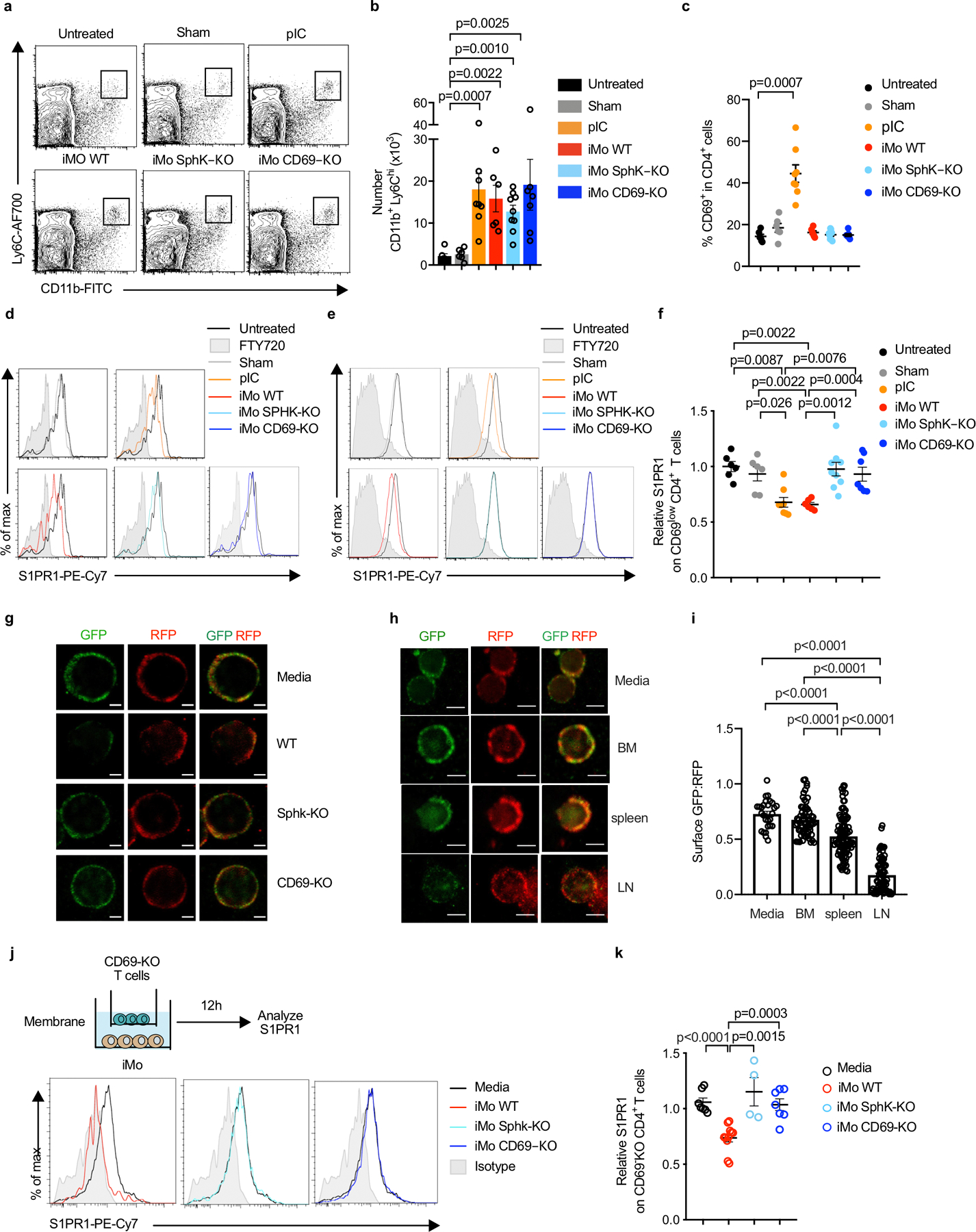
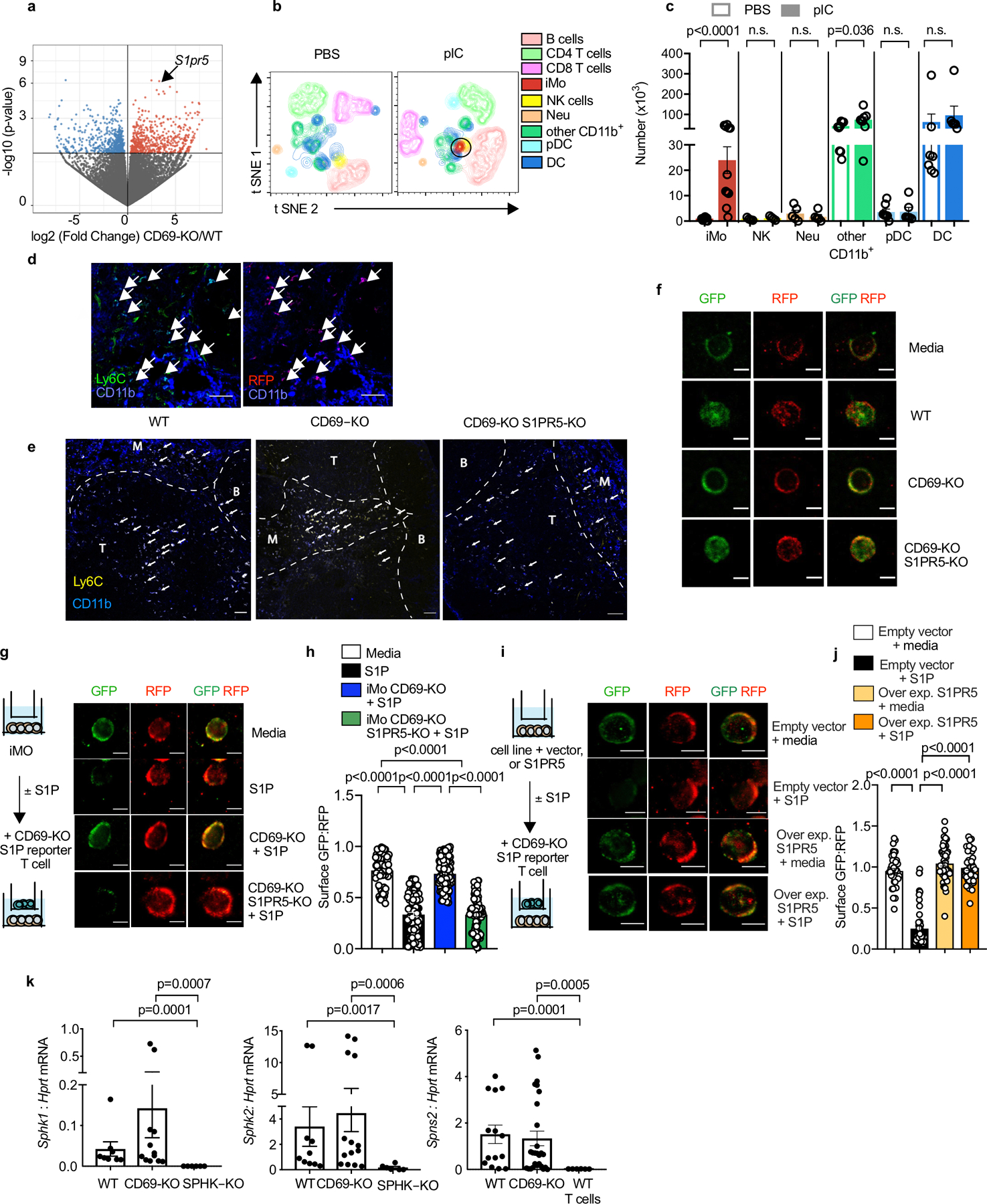
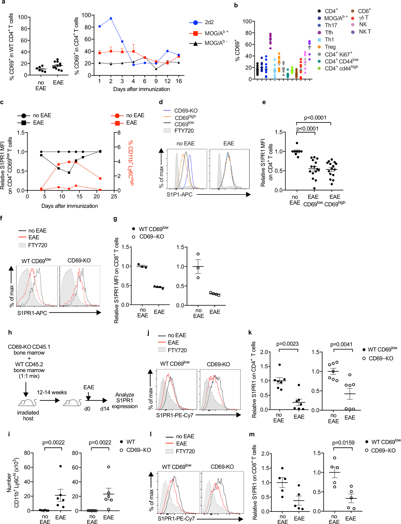
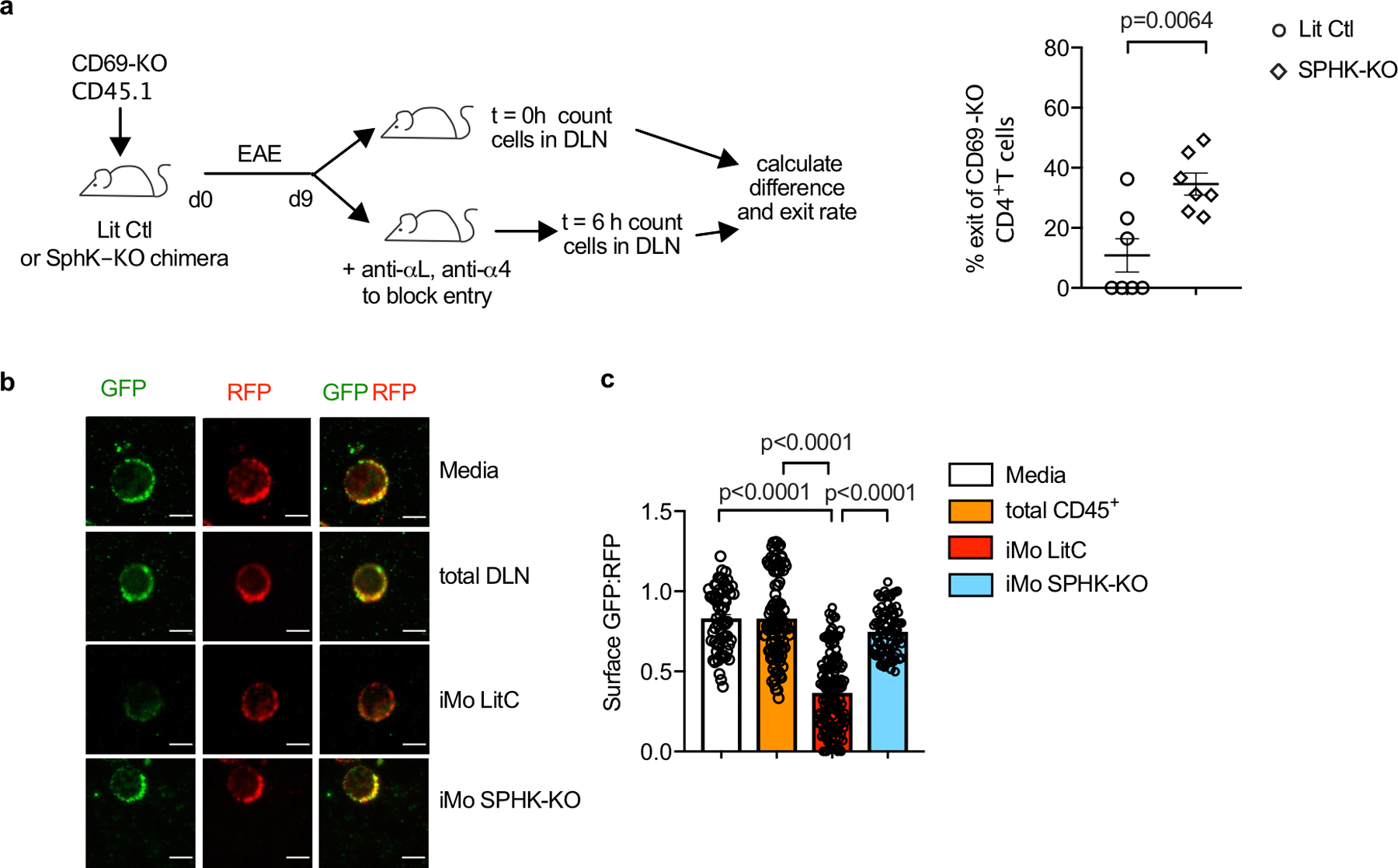
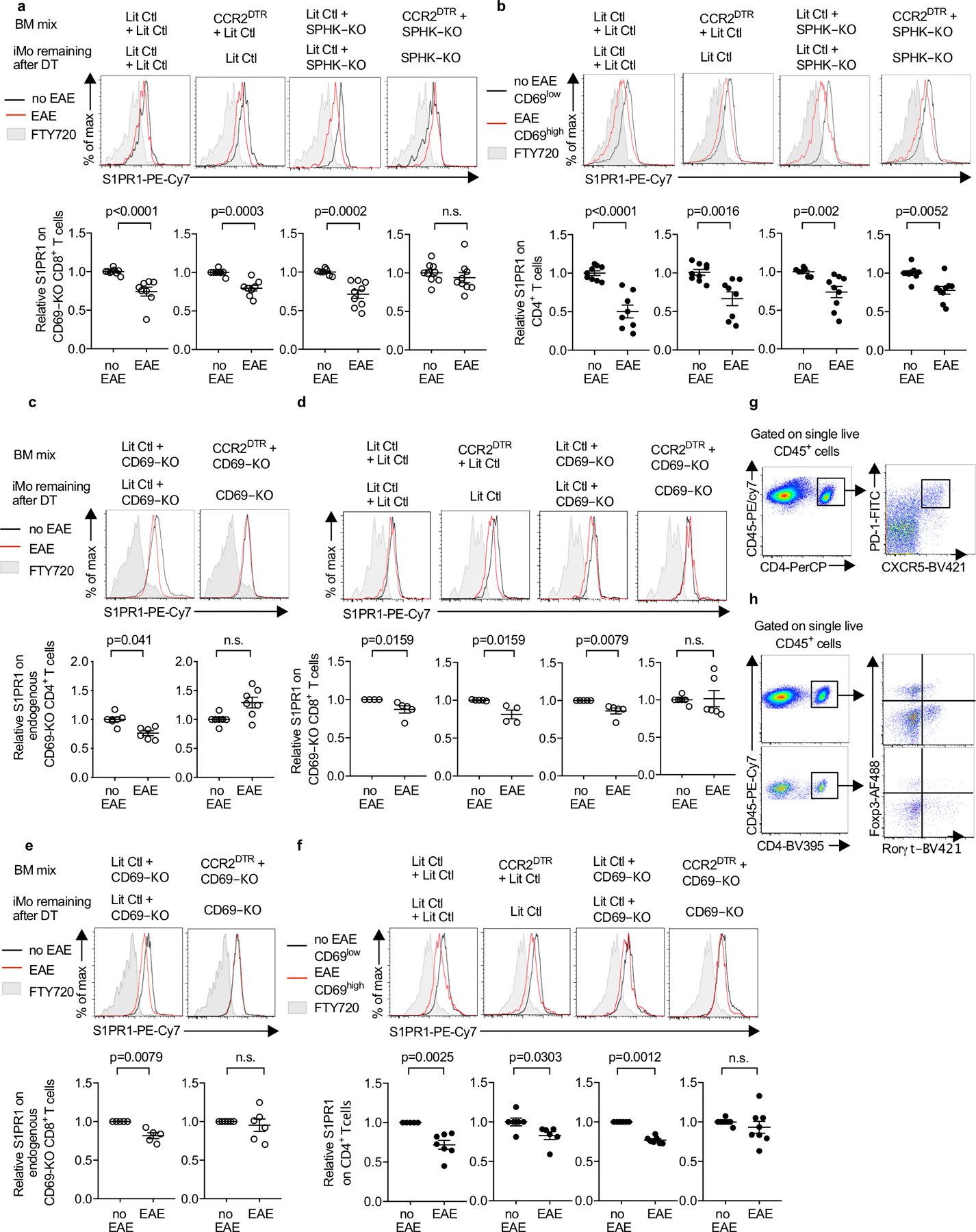
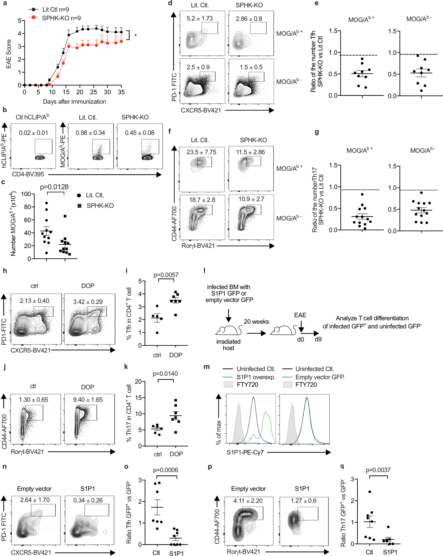
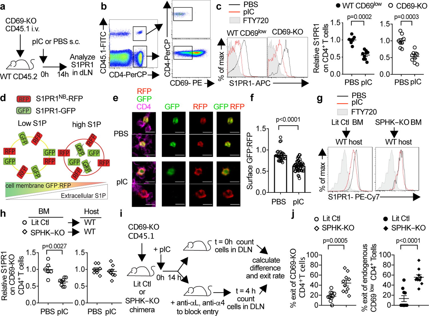
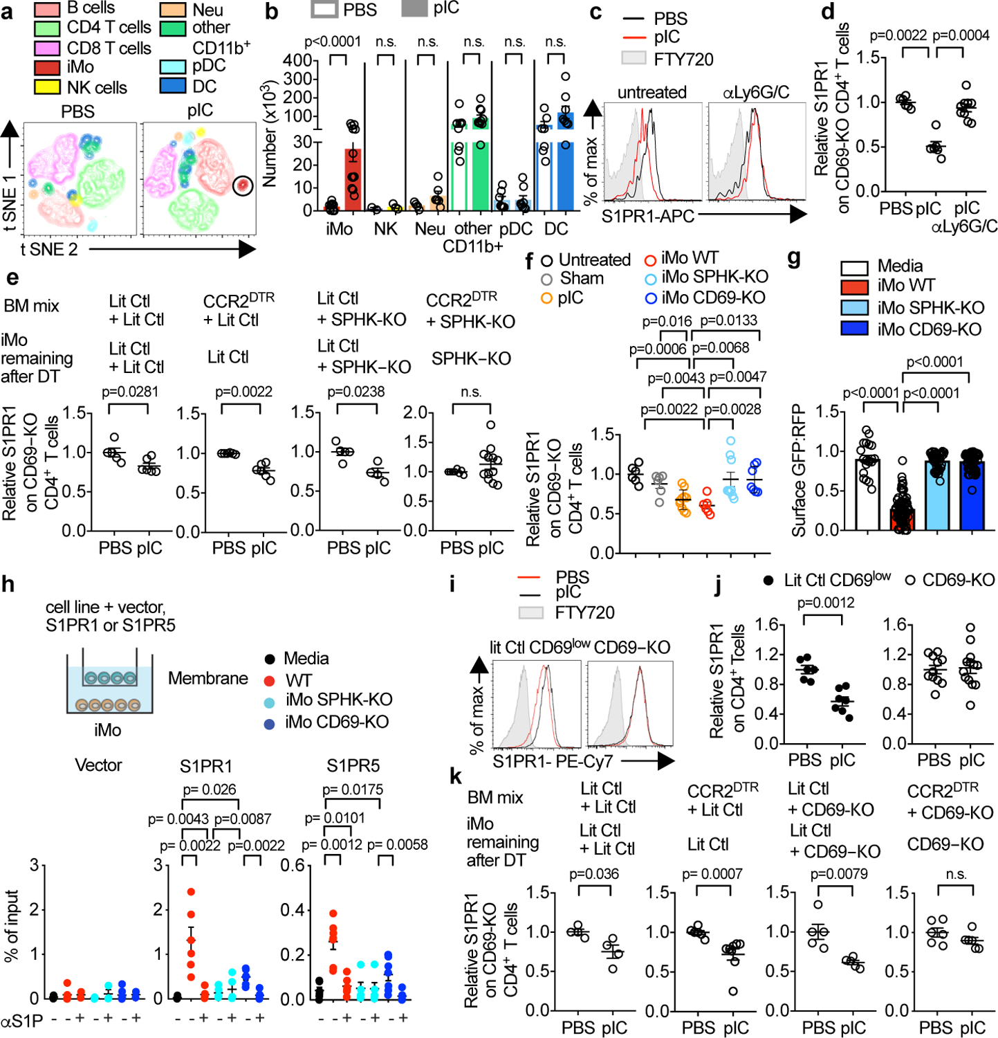
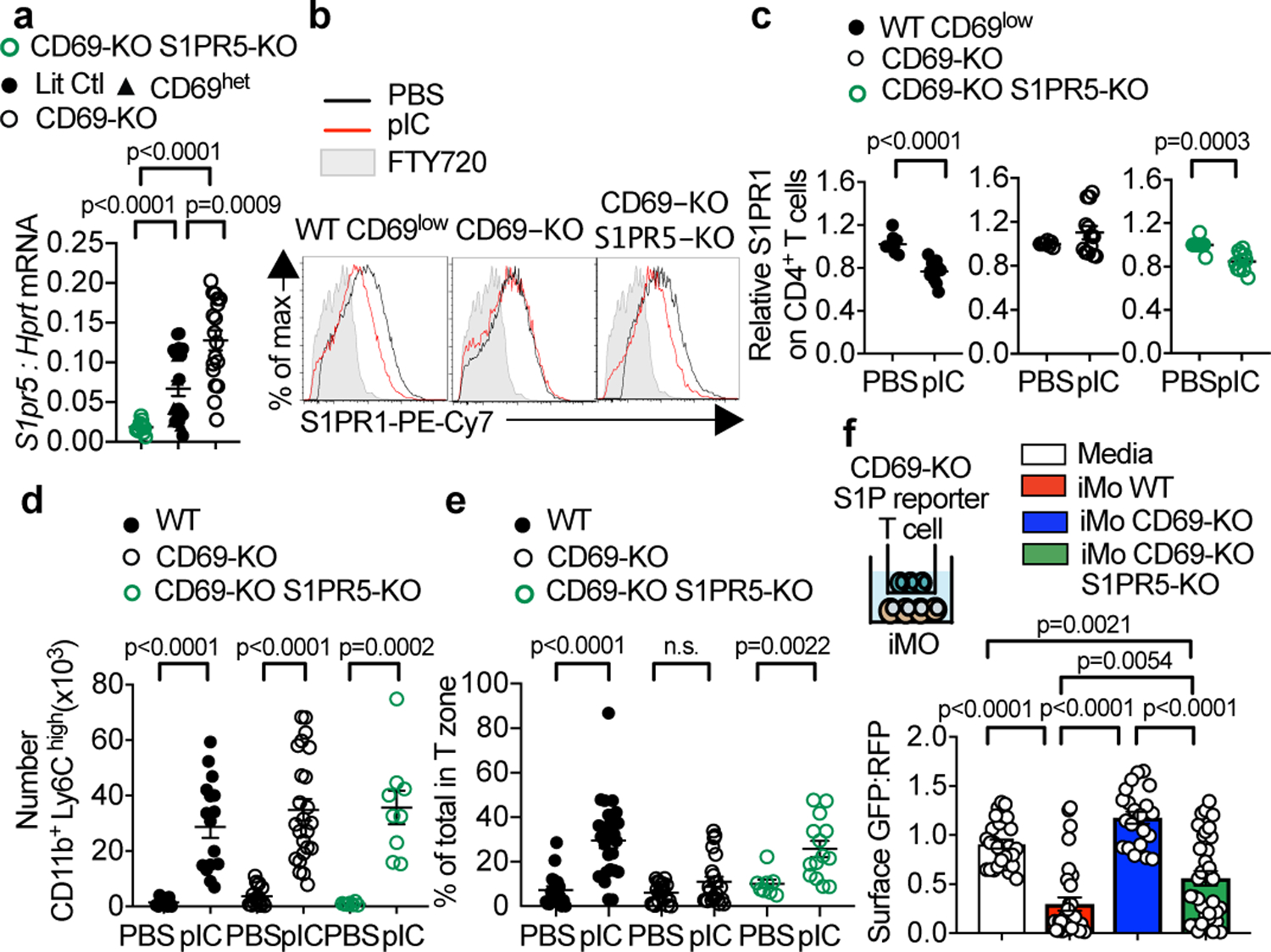
The lipid chemoattractant sphingosine 1-phosphate (S1P) guides cells out of tissues, where the concentration of S1P is relatively low, into circulatory fluids, where the concentration of S1P is high. For example, S1P directs the exit of T cells from lymph nodes, where T cells are initially activated, into lymph, from which T cells reach the blood and ultimately inflamed tissues. T cells follow S1P gradients primarily using S1P receptor 1 (ref. ). Recent studies have described how S1P gradients are established at steady state, but little is known about the distribution of S1P in disease or about how changing levels of S1P may affect immune responses. Here we show that the concentration of S1P increases in lymph nodes during an immune response. We found that haematopoietic cells, including inflammatory monocytes, were an important source of this S1P, which was an unexpected finding as endothelial cells provide S1P to lymph. Inflammatory monocytes required the early activation marker CD69 to supply this S1P, in part because the expression of CD69 was associated with reduced levels of S1pr5 (which encodes S1P receptor 5). CD69 acted as a 'stand-your-ground' signal, keeping immune cells at a site of inflammation by regulating both the receptors and the gradients of S1P. Finally, increased levels of S1P prolonged the residence time of T cells in the lymph nodes and exacerbated the severity of experimental autoimmune encephalomyelitis in mice. This finding suggests that residence time in the lymph nodes might regulate the differentiation of T cells, and points to new uses of drugs that target S1P signalling.
脂质趋化因子 1-磷酸鞘氨醇(S1P)引导细胞离开组织,组织中 S1P 的浓度相对较低,进入循环液,循环液中 S1P 的浓度较高。例如,S1P 指导 T 细胞从最初被激活的淋巴结中排出,进入淋巴液,T 细胞从淋巴液中进入血液,最终进入炎症组织。T 细胞主要使用 S1P 受体 1(参考文献)来跟踪 S1P 梯度。最近的研究描述了 S1P 梯度如何在稳态下建立,但对于 S1P 在疾病中的分布以及 S1P 水平的变化如何影响免疫反应知之甚少。在这里,我们表明在免疫反应过程中,淋巴结中的 S1P 浓度增加。我们发现,包括炎性单核细胞在内的造血细胞是这种 S1P 的重要来源,这是一个意外的发现,因为内皮细胞为淋巴提供 S1P。炎性单核细胞需要早期激活标志物 CD69 来提供这种 S1P,部分原因是 CD69 的表达与 S1pr5(编码 S1P 受体 5)水平降低有关。CD69 充当“坚守阵地”信号,通过调节 S1P 的受体和梯度,使免疫细胞留在炎症部位。最后,S1P 水平的升高延长了 T 细胞在淋巴结中的停留时间,并加重了实验性自身免疫性脑脊髓炎小鼠的严重程度。这一发现表明,在淋巴结中的停留时间可能调节 T 细胞的分化,并为靶向 S1P 信号的药物提供了新的用途。