• 文献检索
  • 文档翻译
  • 深度研究
  • 学术资讯
  • Suppr Zotero 插件Zotero 插件
  • 邀请有礼
  • 套餐&价格
  • 历史记录
应用&插件
Suppr Zotero 插件Zotero 插件浏览器插件Mac 客户端Windows 客户端微信小程序
定价
高级版会员购买积分包购买API积分包
服务
文献检索文档翻译深度研究API 文档MCP 服务
关于我们
关于 Suppr公司介绍联系我们用户协议隐私条款
关注我们

Suppr 超能文献

核心技术专利:CN118964589B侵权必究
粤ICP备2023148730 号-1Suppr @ 2026

文献检索

告别复杂PubMed语法,用中文像聊天一样搜索,搜遍4000万医学文献。AI智能推荐,让科研检索更轻松。

立即免费搜索

文件翻译

保留排版,准确专业,支持PDF/Word/PPT等文件格式,支持 12+语言互译。

免费翻译文档

深度研究

AI帮你快速写综述,25分钟生成高质量综述,智能提取关键信息,辅助科研写作。

立即免费体验

胱氨酸病中半胱氨酸晶体的无创性皮内成像。

Non-invasive intradermal imaging of cystine crystals in cystinosis.

机构信息

Division of Genetics, Department of Pediatrics, University of California, San Diego, La Jolla, California, United States of America.

Biostatistics Research Center, Herbert Wertheim School of Public Health and Human Longevity Science, University of California, San Diego, La Jolla, California, United States of America.

出版信息

PLoS One. 2021 Mar 4;16(3):e0247846. doi: 10.1371/journal.pone.0247846. eCollection 2021.

DOI:10.1371/journal.pone.0247846
PMID:33661986
原文链接:https://pmc.ncbi.nlm.nih.gov/articles/PMC7932553/
Abstract

IMPORTANCE

Development of noninvasive methodology to reproducibly measure tissue cystine crystal load to assess disease status and guide clinical care in cystinosis, an inherited lysosomal storage disorder characterized by widespread cystine crystal accumulation.

OBJECTIVE

To develop an unbiased and semi-automated imaging methodology to quantify dermal cystine crystal accumulation in patients to correlate with disease status.

DESIGN, SETTING AND PARTICIPANTS: 101 participants, 70 patients and 31 healthy controls, were enrolled at the University of California, San Diego, Cystinosis Clinics, Rady Children's Hospital, San Diego and at the annual Cystinosis Research Foundation family conference for an ongoing prospective longitudinal cohort study of cystinosis patients with potential yearly follow-up.

EXPOSURES

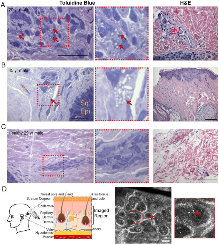
Intradermal reflectance confocal microscopy (RCM) imaging, blood collection via standard venipuncture, medical record collection, and occasional skin punch biopsies.

MAIN OUTCOMES AND MEASURES

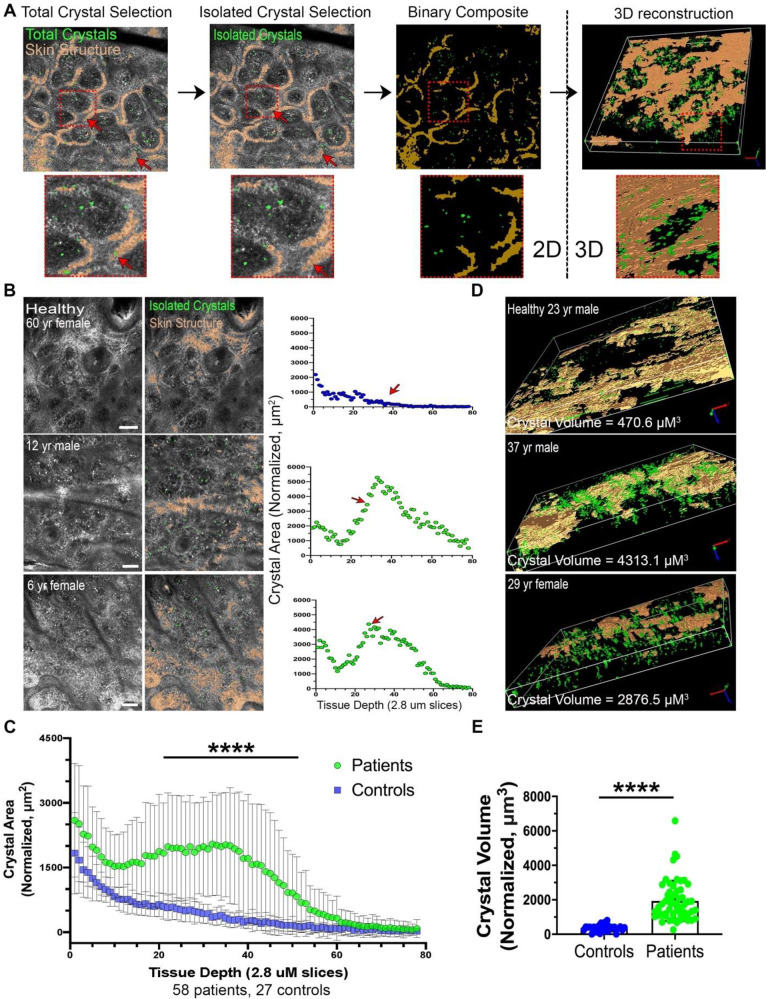
The primary outcome was to establish an automated measure of normalized confocal crystal volume (nCCV) for each subject. Secondary analysis examined the association of nCCV with various clinical indicators to assess nCCV's possible predictive potential.

RESULTS

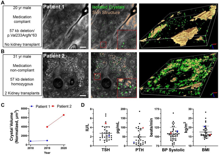
Over 2 years, 57 patients diagnosed with cystinosis (median [range] age: 15.1 yrs [0.8, 54]; 41.4% female) were intradermally assessed by RCM to produce 84 image stacks. 27 healthy individuals (38.7 yrs [10, 85]; 53.1% female) were also imaged providing 37 control image stacks. Automated 2D crystal area quantification revealed that patients had significantly elevated crystal accumulation within the superficial dermis. 3D volumetric analysis of this region was significantly higher in patients compared to healthy controls (mean [SD]: 1934.0 μm3 [1169.1] for patients vs. 363.1 μm3 [194.3] for controls, P<0.001). Medical outcome data was collected from 43 patients with infantile cystinosis (media [range] age: 11 yrs [0.8, 54]; 51% female). nCCV was positively associated with hypothyroidism (OR = 19.68, 95% CI: [1.60, 242.46], P = 0.02) and stage of chronic kidney disease (slope estimate = 0.53, 95%CI: [0.05, 1.00], P = 0.03).

CONCLUSIONS AND RELEVANCE

This study used non-invasive RCM imaging to develop an intradermal cystine crystal quantification method. Results showed that cystinosis patients had increased nCCV compared to healthy controls. Level of patient nCCV correlated with several clinical outcomes suggesting nCCV may be used as a potential new biomarker for cystinosis to monitor long-term disease control and medication compliance.

摘要

重要性

开发一种非侵入性的方法来重现性地测量组织胱氨酸晶体负荷,以评估疾病状态并指导胱氨酸病的临床护理,胱氨酸病是一种遗传性溶酶体贮积症,其特征是广泛的胱氨酸晶体积累。

目的

开发一种无偏倚和半自动化的成像方法来定量评估患者皮肤中的胱氨酸晶体积累,以与疾病状态相关联。

设计、设置和参与者:在加利福尼亚大学圣地亚哥分校、圣地亚哥 Rady 儿童医院的胱氨酸病诊所,以及胱氨酸病研究基金会的年度家庭会议上,共招募了 101 名参与者,其中 70 名患者和 31 名健康对照者,他们参与了一项正在进行的前瞻性纵向队列研究,对胱氨酸病患者进行潜在的年度随访。

暴露

皮肤内反射共聚焦显微镜(RCM)成像、通过标准静脉穿刺采集血液、病历采集和偶尔的皮肤打孔活检。

主要结果和测量指标

主要结果是为每个受试者建立一个正常化的共聚焦晶体体积(nCCV)的自动测量方法。二级分析检查了 nCCV 与各种临床指标的关联,以评估 nCCV 的潜在预测潜力。

结果

在 2 年的时间里,对 57 名确诊的胱氨酸病患者(中位数[范围]年龄:15.1 岁[0.8,54];41.4%为女性)进行了 RCM 皮内评估,产生了 84 个图像堆栈。还对 27 名健康个体(38.7 岁[10,85];53.1%为女性)进行了成像,提供了 37 个对照图像堆栈。自动化的 2D 晶体面积定量显示,患者的浅层真皮中晶体积累明显升高。与健康对照组相比,该区域的 3D 体积分析明显更高(患者的平均[标准差]:1934.0 μm3[1169.1],健康对照组为 363.1 μm3[194.3],P<0.001)。对 43 名患有婴儿胱氨酸病的患者(中位数[范围]年龄:11 岁[0.8,54];51%为女性)收集了 nCCV 数据。nCCV 与甲状腺功能减退症呈正相关(比值比[OR]=19.68,95%置信区间[CI]:[1.60,242.46],P=0.02)和慢性肾脏病的分期(斜率估计值=0.53,95%CI:[0.05,1.00],P=0.03)。

结论和相关性

本研究使用非侵入性的 RCM 成像技术开发了一种皮内胱氨酸晶体定量方法。结果表明,胱氨酸病患者的 nCCV 高于健康对照组。患者的 nCCV 水平与多种临床结果相关,表明 nCCV 可能作为胱氨酸病的一种潜在新生物标志物,用于监测长期疾病控制和药物依从性。

https://cdn.ncbi.nlm.nih.gov/pmc/blobs/b713/7932553/64c74d593ab9/pone.0247846.g003.jpg
https://cdn.ncbi.nlm.nih.gov/pmc/blobs/b713/7932553/b4d57165dd50/pone.0247846.g001.jpg
https://cdn.ncbi.nlm.nih.gov/pmc/blobs/b713/7932553/328544b41386/pone.0247846.g002.jpg
https://cdn.ncbi.nlm.nih.gov/pmc/blobs/b713/7932553/64c74d593ab9/pone.0247846.g003.jpg
https://cdn.ncbi.nlm.nih.gov/pmc/blobs/b713/7932553/b4d57165dd50/pone.0247846.g001.jpg
https://cdn.ncbi.nlm.nih.gov/pmc/blobs/b713/7932553/328544b41386/pone.0247846.g002.jpg
https://cdn.ncbi.nlm.nih.gov/pmc/blobs/b713/7932553/64c74d593ab9/pone.0247846.g003.jpg

相似文献

1
Non-invasive intradermal imaging of cystine crystals in cystinosis.胱氨酸病中半胱氨酸晶体的无创性皮内成像。
PLoS One. 2021 Mar 4;16(3):e0247846. doi: 10.1371/journal.pone.0247846. eCollection 2021.
2
In vivo reflectance confocal microscopy of the skin: a noninvasive means of assessing body cystine accumulation in infantile cystinosis.皮肤共聚焦激光扫描显微镜:一种非侵入性评估婴儿胱氨酸贮积症皮肤胱氨酸蓄积的方法。
J Am Acad Dermatol. 2013 Apr;68(4):e111-e116. doi: 10.1016/j.jaad.2011.08.010. Epub 2011 Oct 2.
3
Spectral domain optical coherence tomography-based retinochoroidal cystine crystal score: a window into infantile nephropathic cystinosis.基于光谱域光学相干断层扫描的视网膜脉络膜胱氨酸晶体评分:洞察婴儿型肾病性胱氨酸病的窗口。
Br J Ophthalmol. 2023 Feb;107(2):234-241. doi: 10.1136/bjophthalmol-2021-319612. Epub 2021 Sep 16.
4
Examination of corneal deposits in nephropathic cystinosis using in vivo confocal microscopy and anterior segment optical coherence tomography: an age-dependent cross sectional study.使用活体共聚焦显微镜和眼前节光学相干断层扫描检查肾病性胱氨酸病中的角膜沉积物:一项年龄相关性横断面研究。
BMC Ophthalmol. 2020 Feb 26;20(1):73. doi: 10.1186/s12886-020-01336-w.
5
Establishing an objective biomarker for corneal cystinosis using a threshold-based Spectral domain optical coherence tomography imaging algorithm.利用基于阈值的谱域光学相干断层扫描成像算法建立角膜胱氨酸沉积症的客观生物标志物。
Acta Ophthalmol. 2021 Mar;99(2):e189-e195. doi: 10.1111/aos.14569. Epub 2020 Aug 24.
6
Clinical utility of chitotriosidase enzyme activity in nephropathic cystinosis.几丁质酶活性在肾病性胱氨酸病中的临床应用
Orphanet J Rare Dis. 2014 Nov 19;9:155. doi: 10.1186/s13023-014-0155-z.
7
A new gel formulation of topical cysteamine for the treatment of corneal cystine crystals in cystinosis: the Cystadrops OCT-1 study.一种用于治疗胱氨酸病中角膜胱氨酸结晶的新型局部用半胱胺凝胶制剂:Cystadrops OCT-1研究。
Mol Genet Metab. 2014 Mar;111(3):314-320. doi: 10.1016/j.ymgme.2013.12.298. Epub 2014 Jan 9.
8
In vivo confocal microscopy and anterior segment optical coherence tomography analysis of the cornea in nephropathic cystinosis.肾病性胱氨酸病患者角膜的体内共聚焦显微镜检查和眼前节光学相干断层扫描分析
Ophthalmology. 2009 May;116(5):870-6. doi: 10.1016/j.ophtha.2008.11.021.
9
Late-onset nephropathic cystinosis: clinical presentation, outcome, and genotyping.迟发性肾病型胱氨酸病:临床表现、预后及基因分型
Clin J Am Soc Nephrol. 2008 Jan;3(1):27-35. doi: 10.2215/CJN.01740407.
10
A potential new method to estimate tissue cystine content in nephropathic cystinosis.一种估计肾性胱氨酸贮积症组织胱氨酸含量的新方法。
J Pediatr. 2012 Sep;161(3):531-535.e1. doi: 10.1016/j.jpeds.2012.03.011. Epub 2012 Apr 17.

引用本文的文献

1
Biomarkers in Nephropathic Cystinosis: Current and Future Perspectives.遗传性胱氨酸病的生物标志物:现状和未来展望。
Cells. 2022 Jun 4;11(11):1839. doi: 10.3390/cells11111839.
2
Hematopoietic Stem Cell Gene Therapy for Cystinosis: From Bench-to-Bedside.胱氨酸病的造血干细胞基因治疗:从基础到临床。
Cells. 2021 Nov 23;10(12):3273. doi: 10.3390/cells10123273.

本文引用的文献

1
Reflectance confocal microscopy and dermoscopy aid in evaluating repigmentation within or adjacent to lentigo maligna melanoma surgical scars.反射式共聚焦显微镜和皮肤镜有助于评估黑素细胞痣恶性黑素瘤手术疤痕内或周围的复色情况。
J Eur Acad Dermatol Venereol. 2020 Jan;34(1):74-81. doi: 10.1111/jdv.15819. Epub 2019 Aug 23.
2
In Vivo Reflectance Confocal Microscopy: Emerging Role in Noninvasive Diagnosis and Monitoring of Eczematous Dermatoses.体内反射共聚焦显微镜检查:在湿疹性皮肤病无创诊断和监测中的新兴作用
Actas Dermosifiliogr (Engl Ed). 2019 Oct;110(8):626-636. doi: 10.1016/j.ad.2018.08.008. Epub 2019 Jun 13.
3
Interaction between galectin-3 and cystinosin uncovers a pathogenic role of inflammation in kidney involvement of cystinosis.
半乳糖凝集素-3 与胱氨酸蛋白酶抑制剂的相互作用揭示了炎症在胱氨酸病肾脏受累中的致病作用。
Kidney Int. 2019 Aug;96(2):350-362. doi: 10.1016/j.kint.2019.01.029. Epub 2019 Mar 6.
4
Nephropathic Cystinosis: Symptoms, Treatment, and Perspectives of a Systemic Disease.肾性胱氨酸病:一种全身性疾病的症状、治疗及展望
Front Pediatr. 2018 Mar 14;6:58. doi: 10.3389/fped.2018.00058. eCollection 2018.
5
Endocrine Complications of Cystinosis.胱氨酸病的内分泌并发症
J Pediatr. 2017 Apr;183S:S5-S8. doi: 10.1016/j.jpeds.2016.12.049.
6
The renal Fanconi syndrome in cystinosis: pathogenic insights and therapeutic perspectives.胱氨酸病中的肾范可尼综合征:发病机制的见解和治疗观点。
Nat Rev Nephrol. 2017 Feb;13(2):115-131. doi: 10.1038/nrneph.2016.182. Epub 2016 Dec 19.
7
Diagnosis and Monitoring of Cystinosis Using Immunomagnetically Purified Granulocytes.采用免疫磁珠纯化粒细胞对胱氨酸病进行诊断和监测。
Clin Chem. 2016 May;62(5):766-72. doi: 10.1373/clinchem.2015.252494. Epub 2016 Mar 15.
8
Cysteamine (Cystagon®) adherence in patients with cystinosis in Spain: successful in children and a challenge in adolescents and adults.西班牙胱氨酸病患者对半胱胺(Cystagon®)的依从性:儿童患者效果良好,青少年及成人患者面临挑战。
Nephrol Dial Transplant. 2015 Mar;30(3):475-80. doi: 10.1093/ndt/gfu329. Epub 2014 Oct 26.
9
Cystinosis: clinical presentation, pathogenesis and treatment.胱氨酸病:临床表现、发病机制与治疗
Pediatr Endocrinol Rev. 2014 Sep;12 Suppl 1:176-84.
10
Age-related morphometric changes of inner structures of the skin assessed by in vivo reflectance confocal microscopy.通过体内反射式共聚焦显微镜评估皮肤内部结构的年龄相关形态计量学变化。
Int J Dermatol. 2015 Mar;54(3):295-301. doi: 10.1111/ijd.12220. Epub 2014 Sep 30.