Department of Orthopedics, The People's Hospital of China Three Gorges University, The First People's Hospital of Yichang, Yichang, Hubei, China.
Department of Orthopedics, The People's Hospital of WuFeng, Yichang, Hubei, China.
Sci Rep. 2021 Mar 4;11(1):5111. doi: 10.1038/s41598-021-84164-2.
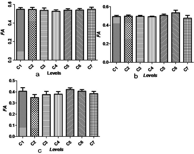
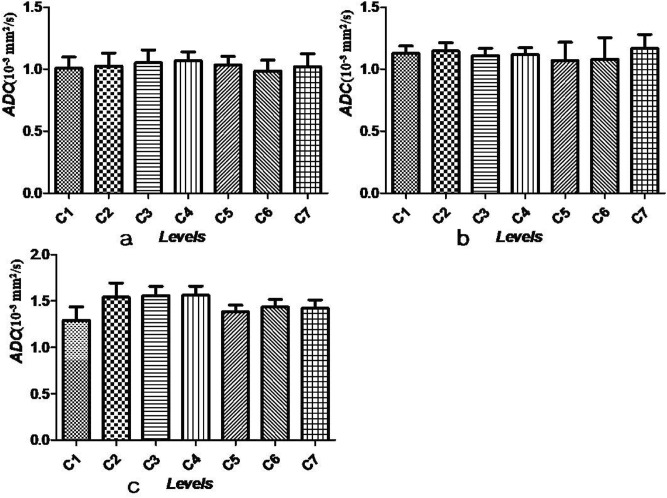
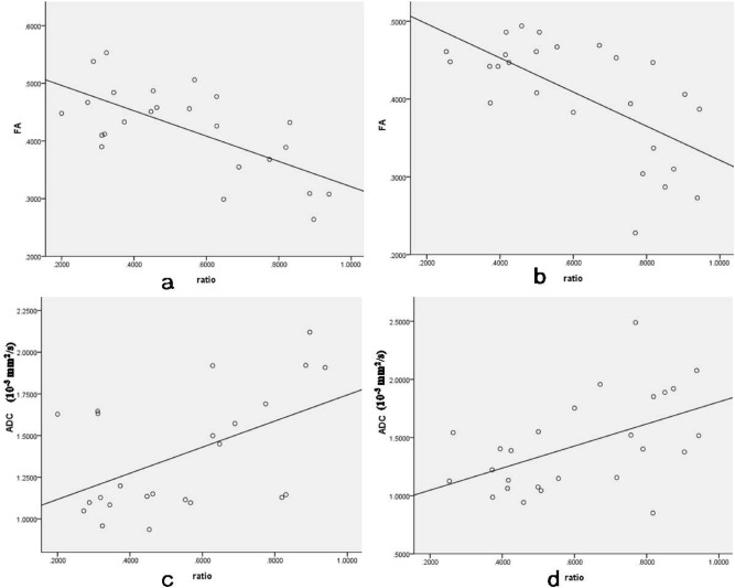
The microstructure of the spinal cord in syringomyelia has not been well studied. The aim of this study was to evaluate the microstructure of the cervical cord in patients with syringomyelia using diffusion tensor imaging (DTI) and to investigate the association between DTI parameters and the size of the syrinx cavity. Thirty patients with syringomyelia and 11 age-matched controls were included in this study. DTI and T1/T2-weighted MRI were used to estimate spinal microstructure. The patients were divided into a clinical symptom group (group A) and a non-clinical symptom group (group B) according to ASIA assessments. The fractional anisotropy (FA) and apparent diffusion coefficient (ADC) values (mm/s) were measured and compared between patients and controls. Correlation between FA/ADC and the size of the syrinx cavity was examined with a bivariate analysis. FA values were lower (P < 0.000) and ADC values were higher (P < 0.000) compared to the controls at the level of all syrinxes examined in patients with syringomyelia; both FA values and ADC values reached normal values either above or below the syrinx levels (all P > 0.05). FA values and ADC values at all cervical levels were not significantly different either in controls or outside of the syrinx (all P > 0.05). FA values of group A was significantly lower than those of group B (P < 0.000). There was a negative association between FA values and the size of syrinx cavity, and a positive association between ADC values and the size of syrinx cavity (FA: P < 0.05, ADC: P < 0.05). The microstructure of the cervical spinal cord is different across all patients with syringomyelia. DTI is a promising tool for estimating quantitative pathological characteristics that are not visible with general MRI.
脊髓空洞症的脊髓微观结构尚未得到很好的研究。本研究旨在使用弥散张量成像(DTI)评估脊髓空洞症患者颈髓的微观结构,并探讨 DTI 参数与空洞腔大小之间的关系。本研究纳入了 30 例脊髓空洞症患者和 11 名年龄匹配的对照者。使用 DTI 和 T1/T2 加权 MRI 来评估脊髓微观结构。根据 ASIA 评估,患者分为临床症状组(A 组)和非临床症状组(B 组)。测量并比较了患者和对照组之间的各向异性分数(FA)和表观扩散系数(ADC)值(mm/s)。用双变量分析检查 FA/ADC 与空洞腔大小之间的相关性。与对照组相比,脊髓空洞症患者所有空洞水平的 FA 值均较低(P<0.000),ADC 值均较高(P<0.000);FA 值和 ADC 值在空洞上下水平均恢复正常(均 P>0.05)。在对照组或空洞外,所有颈椎水平的 FA 值和 ADC 值均无显著差异(均 P>0.05)。A 组的 FA 值明显低于 B 组(P<0.000)。FA 值与空洞腔大小呈负相关,ADC 值与空洞腔大小呈正相关(FA:P<0.05,ADC:P<0.05)。所有脊髓空洞症患者的颈髓微观结构均不同。DTI 是一种很有前途的工具,可用于评估常规 MRI 无法显示的定量病理特征。