Cepeda Santiago, García-García Sergio, Velasco-Casares María, Fernández-Pérez Gabriel, Zamora Tomás, Arrese Ignacio, Sarabia Rosario
Department of Neurosurgery, University Hospital Río Hortega, 47012 Valladolid, Spain.
Department of Radiology, University Hospital Río Hortega, 47012 Valladolid, Spain.
Brain Sci. 2021 Feb 21;11(2):271. doi: 10.3390/brainsci11020271.
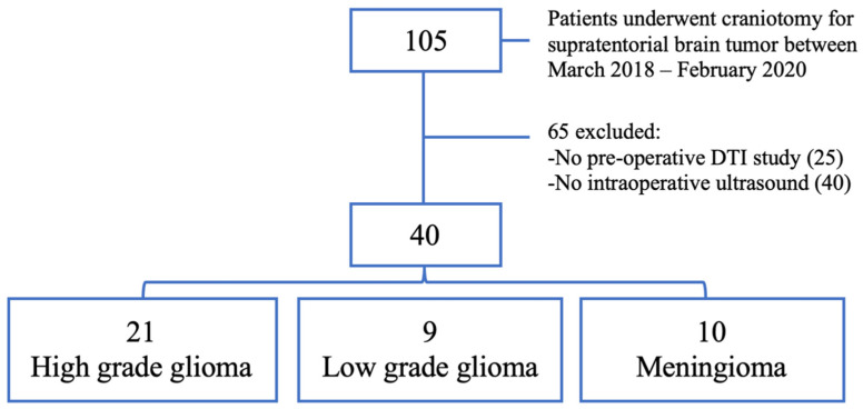
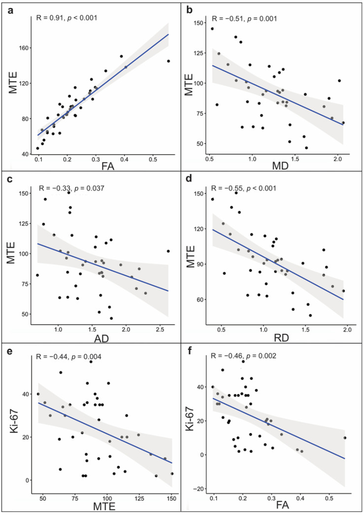
Intraoperative ultrasound elastography (IOUS-E) is a novel image modality applied in brain tumor assessment. However, the potential links between elastographic findings and other histological and neuroimaging features are unknown. This study aims to find associations between brain tumor elasticity, diffusion tensor imaging (DTI) metrics, and cell proliferation. A retrospective study was conducted to analyze consecutively admitted patients who underwent craniotomy for supratentorial brain tumors between March 2018 and February 2020. Patients evaluated by IOUS-E and preoperative DTI were included. A semi-quantitative analysis was performed to calculate the mean tissue elasticity (MTE). Diffusion coefficients and the tumor proliferation index by Ki-67 were registered. Relationships between the continuous variables were determined using the Spearman ρ test. A predictive model was developed based on non-linear regression using the MTE as the dependent variable. Forty patients were evaluated. The pathologic diagnoses were as follows: 21 high-grade gliomas (HGG); 9 low-grade gliomas (LGG); and 10 meningiomas. Cases with a proliferation index of less than 10% had significantly higher medians of MTE (110.34 vs. 79.99, < 0.001) and fractional anisotropy (FA) (0.24 vs. 0.19, = 0.020). We found a strong positive correlation between MTE and FA ( (38) = 0.91, < 0.001). A cubic spline non-linear regression model was obtained to predict tumoral MTE from FA ( = 0.78, < 0.001). According to our results, tumor elasticity is associated with histopathological and DTI-derived metrics. These findings support the usefulness of IOUS-E as a complementary tool in brain tumor surgery.
术中超声弹性成像(IOUS-E)是一种应用于脑肿瘤评估的新型成像方式。然而,弹性成像结果与其他组织学和神经影像学特征之间的潜在联系尚不清楚。本研究旨在寻找脑肿瘤弹性、扩散张量成像(DTI)指标与细胞增殖之间的关联。进行了一项回顾性研究,以分析2018年3月至2020年2月期间因幕上脑肿瘤接受开颅手术的连续入院患者。纳入接受IOUS-E评估和术前DTI的患者。进行半定量分析以计算平均组织弹性(MTE)。记录扩散系数和Ki-67的肿瘤增殖指数。使用Spearman ρ检验确定连续变量之间的关系。以MTE作为因变量,基于非线性回归建立预测模型。评估了40例患者。病理诊断如下:21例高级别胶质瘤(HGG);9例低级别胶质瘤(LGG);10例脑膜瘤。增殖指数小于10%的病例MTE中位数(110.34对79.99,<0.001)和各向异性分数(FA)(0.24对0.19,=0.020)显著更高。我们发现MTE与FA之间存在强正相关((38)=0.91,<0.001)。获得了一个三次样条非线性回归模型,用于从FA预测肿瘤MTE(=0.78,<0.001)。根据我们的结果,肿瘤弹性与组织病理学和DTI衍生指标相关。这些发现支持IOUS-E作为脑肿瘤手术中一种辅助工具的实用性。