Department of Medicine and Surgery, University of Parma, 43100 Parma, Italy.
Cognitive and Motor Center, Medicine and Geriatric-Rehabilitation Department of Parma, University-Hospital of Parma, 43126 Parma, Italy.
Int J Environ Res Public Health. 2021 Feb 28;18(5):2356. doi: 10.3390/ijerph18052356.
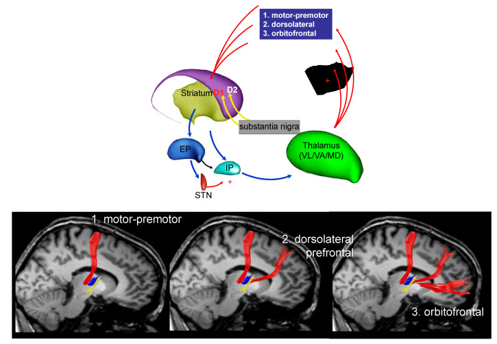
The neurobiology of Parkinson's disease and its progression has been investigated during the last few decades. Braak et al. proposed neuropathological stages of this disease based on the recognizable topographical extent of Lewy body lesions. This pathological process involves specific brain areas with an ascending course from the brain stem to the cortex. Post-mortem studies are of importance to better understand not only the progression of motor symptoms, but also the involvement of other domains, including cognition and behavior. The correlation between the neuropathological expansion of the disease and the clinical phases remains demanding. Neuroimaging, including magnetic resonance imaging (MRI), positron emission tomography (PET), and single photon emission computed tomography (SPECT), could help to bridge this existing gap by providing in vivo evidence of the extension of the disorders. In the last decade, we observed an overabundance of reports regarding the sensitivity of neuroimaging techniques. All these studies were aimed at improving the accuracy of Parkinson's disease (PD) diagnosis and discriminating it from other causes of parkinsonism. In this review, we look at the recent literature concerning PD and address the new frontier of diagnostic accuracy in terms of identification of early stages of the disease and conventional neuroimaging techniques that, in vivo, are capable of photographing the basal ganglia network and its cerebral connections.
在过去的几十年里,人们一直在研究帕金森病的神经生物学及其进展。Braak 等人根据路易体病变的可识别拓扑范围提出了这种疾病的神经病理学阶段。这个病理过程涉及到特定的大脑区域,其病变从脑干呈上升趋势发展到大脑皮层。尸检研究对于更好地理解运动症状的进展以及其他领域的涉及,包括认知和行为,非常重要。疾病的神经病理学扩展与临床阶段之间的相关性仍然具有挑战性。神经影像学,包括磁共振成像(MRI)、正电子发射断层扫描(PET)和单光子发射计算机断层扫描(SPECT),可以通过提供疾病扩展的活体证据来帮助弥合这一现有差距。在过去的十年中,我们观察到大量关于神经影像学技术敏感性的报告。所有这些研究的目的都是提高帕金森病(PD)诊断的准确性,并将其与其他帕金森综合征的原因区分开来。在这篇综述中,我们回顾了最近关于 PD 的文献,并探讨了诊断准确性的新前沿,即识别疾病的早期阶段,以及能够在体内拍摄基底神经节网络及其大脑连接的常规神经影像学技术。