• 文献检索
  • 文档翻译
  • 深度研究
  • 学术资讯
  • Suppr Zotero 插件Zotero 插件
  • 邀请有礼
  • 套餐&价格
  • 历史记录
应用&插件
Suppr Zotero 插件Zotero 插件浏览器插件Mac 客户端Windows 客户端微信小程序
定价
高级版会员购买积分包购买API积分包
服务
文献检索文档翻译深度研究API 文档MCP 服务
关于我们
关于 Suppr公司介绍联系我们用户协议隐私条款
关注我们

Suppr 超能文献

核心技术专利:CN118964589B侵权必究
粤ICP备2023148730 号-1Suppr @ 2026

文献检索

告别复杂PubMed语法,用中文像聊天一样搜索,搜遍4000万医学文献。AI智能推荐,让科研检索更轻松。

立即免费搜索

文件翻译

保留排版,准确专业,支持PDF/Word/PPT等文件格式,支持 12+语言互译。

免费翻译文档

深度研究

AI帮你快速写综述,25分钟生成高质量综述,智能提取关键信息,辅助科研写作。

立即免费体验

施莱姆管扩张的影响:生物力学及微侵袭性青光眼手术的意义

Effects of Schlemm's Canal Expansion: Biomechanics and MIGS Implications.

作者信息

Xin Chen, Song Shaozhen, Wang Ningli, Wang Ruikang, Johnstone Murray

机构信息

Beijing Tongren Eye Center, Beijing Institute of Ophthalmology, Beijing Tongren Hospital, Capital Medical University, Beijing 100730, China.

Department of Bioengineering, University of Washington, Seattle, WA 98195, USA.

出版信息

Life (Basel). 2021 Feb 23;11(2):176. doi: 10.3390/life11020176.

DOI:10.3390/life11020176
PMID:33672433
原文链接:https://pmc.ncbi.nlm.nih.gov/articles/PMC7926642/
Abstract

OBJECTIVE

To evaluate the change of biomechanical properties of the trabecular meshwork (TM) and configuration of collector channels (CC) by high-resolution optical coherence tomography (HR-OCT) induced by Schlemm's canal (SC) dilation.

METHODS

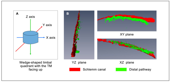
The anterior segments of two human eyes were divided into four quadrants. One end of a specially designed cannula was placed in SC and the other end connected to a perfusion reservoir. HR-OCT provided three-dimensional (3D) volumetric and two-dimensional (2D) cross-sectional imaging permitting assessment of the biomechanical properties of the TM. A large fluid bolus was introduced into SC. Same-sample, pre and post deformation and disruption of SC and CC lumen areas were analyzed.

RESULTS

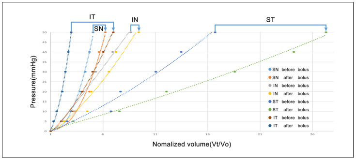
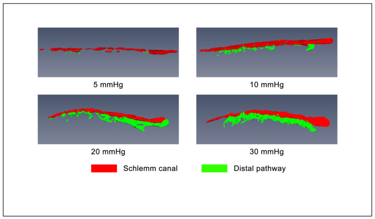
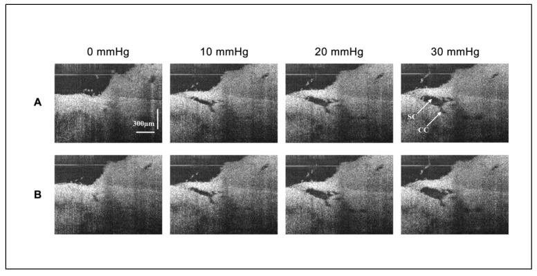
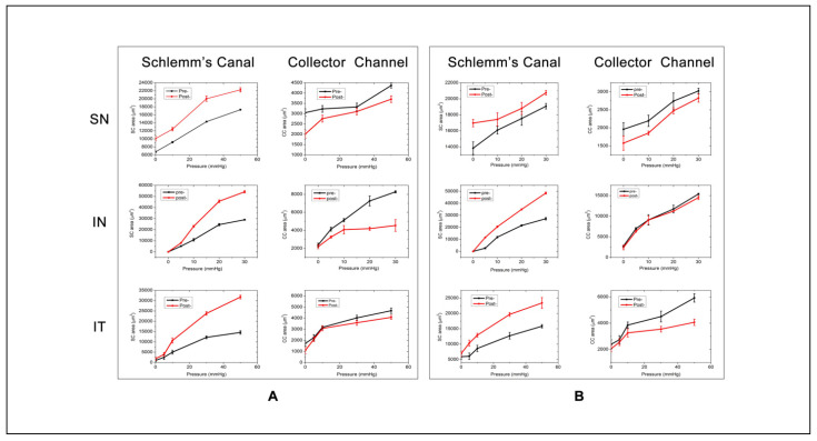
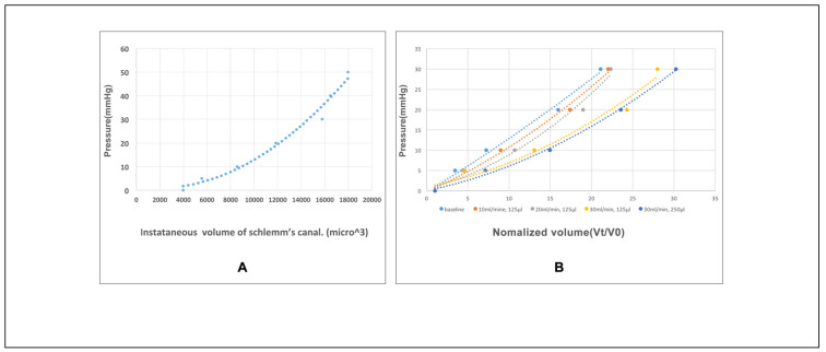
Morphologic 3D reconstructions documented pressure-dependent changes in lumen dimension of SC, CC, and circumferential intrascleral channels. 2D imaging established volumetric stress-strain curves (elastance curves) of the TM in quadrants. The curves of TM elastance shift to the right with an increase in pressure-dependent steady-state SC area. After a bolus disruption, the SC area increased, while the CC area decreased.

CONCLUSION

Our experimental setup permits the study of the biomechanical properties of TM by examining elastance, which differs segmentally and is altered by mechanical expansion of SC by a fluid bolus. The study may shed light on mechanisms of intraocular pressure control of some glaucoma surgery.

摘要

目的

通过高分辨率光学相干断层扫描(HR-OCT)评估施莱姆管(SC)扩张引起的小梁网(TM)生物力学特性变化及集液管(CC)形态。

方法

将两只人眼的眼前节分为四个象限。将一根特制套管的一端置于SC内,另一端连接至灌注储液器。HR-OCT提供三维(3D)容积成像和二维(2D)断层成像,用于评估TM的生物力学特性。向SC内注入大量液体。分析同一标本在SC和CC管腔区域变形和破坏前后的情况。

结果

形态学3D重建记录了SC、CC和巩膜内周向通道管腔尺寸的压力依赖性变化。2D成像建立了各象限TM的容积应力-应变曲线(弹性曲线)。随着压力依赖性稳态SC面积增加,TM弹性曲线右移。注入液体破坏后,SC面积增加,而CC面积减小。

结论

我们的实验装置通过检测弹性来研究TM的生物力学特性,弹性在各节段有所不同,并因液体注入导致的SC机械扩张而改变。该研究可能有助于揭示某些青光眼手术眼压控制的机制。

https://cdn.ncbi.nlm.nih.gov/pmc/blobs/56b8/7926642/b61c7348efec/life-11-00176-g006.jpg
https://cdn.ncbi.nlm.nih.gov/pmc/blobs/56b8/7926642/12cf0de392a0/life-11-00176-g001.jpg
https://cdn.ncbi.nlm.nih.gov/pmc/blobs/56b8/7926642/42a5a9c1e722/life-11-00176-g002.jpg
https://cdn.ncbi.nlm.nih.gov/pmc/blobs/56b8/7926642/8d89814dbf51/life-11-00176-g003.jpg
https://cdn.ncbi.nlm.nih.gov/pmc/blobs/56b8/7926642/39f5a8df57ce/life-11-00176-g004.jpg
https://cdn.ncbi.nlm.nih.gov/pmc/blobs/56b8/7926642/93fe9df156b4/life-11-00176-g005.jpg
https://cdn.ncbi.nlm.nih.gov/pmc/blobs/56b8/7926642/b61c7348efec/life-11-00176-g006.jpg

相似文献

1
Effects of Schlemm's Canal Expansion: Biomechanics and MIGS Implications.施莱姆管扩张的影响:生物力学及微侵袭性青光眼手术的意义
Life (Basel). 2021 Feb 23;11(2):176. doi: 10.3390/life11020176.
2
OCT Study of Mechanical Properties Associated with Trabecular Meshwork and Collector Channel Motion in Human Eyes.人眼小梁网和集合管运动相关力学特性的光学相干断层扫描研究
PLoS One. 2016 Sep 6;11(9):e0162048. doi: 10.1371/journal.pone.0162048. eCollection 2016.
3
Valve-Like Outflow System Behavior With Motion Slowing in Glaucoma Eyes: Findings Using a Minimally Invasive Glaucoma Surgery-MIGS-Like Platform and Optical Coherence Tomography Imaging.青光眼眼中伴有运动减慢的瓣膜样流出系统行为:使用微创青光眼手术(MIGS)样平台和光学相干断层扫描成像的研究结果
Front Med (Lausanne). 2022 Apr 29;9:815866. doi: 10.3389/fmed.2022.815866. eCollection 2022.
4
Anatomic changes in Schlemm's canal and collector channels in normal and primary open-angle glaucoma eyes using low and high perfusion pressures.在低和高灌注压下使用正常和原发性开角型青光眼眼的施莱姆氏管和收集管的解剖学变化。
Invest Ophthalmol Vis Sci. 2014 Aug 19;55(9):5834-41. doi: 10.1167/iovs.14-14128.
5
Biomechanics of human trabecular meshwork in healthy and glaucoma eyes via dynamic Schlemm's canal pressurization.通过动态施莱姆氏管加压研究健康眼和青光眼眼中的人眼小梁网的生物力学。
Comput Methods Programs Biomed. 2022 Jun;221:106921. doi: 10.1016/j.cmpb.2022.106921. Epub 2022 May 27.
6
Effect of age on the morphologies of the human Schlemm's canal and trabecular meshwork measured with swept‑source optical coherence tomography.年龄对使用扫频源光学相干断层扫描测量的人施莱姆管和小梁网形态的影响。
Eye (Lond). 2018 Oct;32(10):1621-1628. doi: 10.1038/s41433-018-0148-6. Epub 2018 Jun 19.
7
Identification and Quantitative Assessment of Schlemm's Canal in the Eyes with 360° Angle Recession Glaucoma.360°房角后退性青光眼眼中施莱姆管的识别与定量评估
J Curr Glaucoma Pract. 2020 Jan-Apr;14(1):25-29. doi: 10.5005/jp-journals-10078-1272.
8
Effects of a Schlemm canal scaffold on collector channel ostia in human anterior segments.施莱姆管支架对人眼前节集合管开口的影响。
Exp Eye Res. 2014 Feb;119:70-6. doi: 10.1016/j.exer.2013.12.011. Epub 2013 Dec 24.
9
Schlemm's canal and trabecular meshwork morphology in high myopia.高度近视眼中的施莱姆管和小梁网形态
Ophthalmic Physiol Opt. 2018 May;38(3):266-272. doi: 10.1111/opo.12451.
10
Novel device for expanding Schlemm's canal: a morphological study: Implantation of the Stegmann Canal Expander following viscodilation of Schlemm's canal resulted in permanent expansion of the canal lumen and distension of the trabecular meshwork without complications.用于扩张施莱姆管的新型装置:一项形态学研究:在对施莱姆管进行粘弹扩张后植入施特格曼管扩张器,可使管腔永久性扩张,并使小梁网扩张,且无并发症。
Br J Ophthalmol. 2015 Jul;99(7):875-7. doi: 10.1136/bjophthalmol-2014-305540. Epub 2015 Jan 18.

引用本文的文献

1
Visual Function After Schlemm's Canal-Based MIGS.基于施莱姆管的微创青光眼手术(MIGS)后的视觉功能
J Clin Med. 2025 Apr 7;14(7):2531. doi: 10.3390/jcm14072531.
2
Two-Year Retrospective Study of Cataract Surgery and Schlemm Canal Hydrus Microstent in Black and Afro-Latino Patients.黑人及拉丁裔黑人患者白内障手术与施累姆管Hydrus微支架的两年回顾性研究
Clin Ophthalmol. 2024 Oct 9;18:2843-2849. doi: 10.2147/OPTH.S481823. eCollection 2024.
3
Historical and Contemporary Debates in Schlemm's Canal-Based MIGS.基于施莱姆管的微切口白内障手术的历史与当代争论

本文引用的文献

1
Reduced Pulsatile Trabecular Meshwork Motion in Eyes With Primary Open Angle Glaucoma Using Phase-Sensitive Optical Coherence Tomography.应用相敏光相干断层扫描技术观察原发性开角型青光眼眼小梁网搏动运动减少。
Invest Ophthalmol Vis Sci. 2020 Dec 1;61(14):21. doi: 10.1167/iovs.61.14.21.
2
Aqueous outflow regulation - 21st century concepts.房水流出调节 - 21 世纪的概念。
Prog Retin Eye Res. 2021 Jul;83:100917. doi: 10.1016/j.preteyeres.2020.100917. Epub 2020 Nov 17.
3
Aqueous outflow imaging techniques and what they tell us about intraocular pressure regulation.
J Clin Med. 2024 Aug 19;13(16):4882. doi: 10.3390/jcm13164882.
4
A Novel Technique Identifies Valve-Like Pathways Entering and Exiting Schlemm's Canal in Primates With Similarities to Human Pathways.一种新技术识别出灵长类动物中进入和离开施莱姆管的瓣膜样通道,这些通道与人类通道相似。
Front Cell Dev Biol. 2022 Jul 4;10:868029. doi: 10.3389/fcell.2022.868029. eCollection 2022.
5
Evaluation of neural innervation in the human conventional outflow pathway distal to Schlemm's canal.评估人类常规流出道中施莱姆管远端的神经支配。
Exp Eye Res. 2022 Aug;221:109132. doi: 10.1016/j.exer.2022.109132. Epub 2022 May 27.
6
Gonioscopy-assisted Transluminal Trabeculotomy (GATT) combined phacoemulsification surgery: Outcomes at a 2-year follow-up.超声乳化白内障吸除术联合房角镜辅助经巩膜隧道小梁切开术:2 年随访结果。
Eye (Lond). 2023 Apr;37(6):1258-1263. doi: 10.1038/s41433-022-02087-2. Epub 2022 May 24.
7
Valve-Like Outflow System Behavior With Motion Slowing in Glaucoma Eyes: Findings Using a Minimally Invasive Glaucoma Surgery-MIGS-Like Platform and Optical Coherence Tomography Imaging.青光眼眼中伴有运动减慢的瓣膜样流出系统行为:使用微创青光眼手术(MIGS)样平台和光学相干断层扫描成像的研究结果
Front Med (Lausanne). 2022 Apr 29;9:815866. doi: 10.3389/fmed.2022.815866. eCollection 2022.
8
Evaluation of different OCT systems in quantitative imaging of human Schlemm's canal.评估不同 OCT 系统在人氏 Schlemm 管定量成像中的应用。
Sci Rep. 2022 Jan 26;12(1):1400. doi: 10.1038/s41598-022-05410-9.
房水流出成像技术及其对眼压调节的启示。
Eye (Lond). 2021 Jan;35(1):216-235. doi: 10.1038/s41433-020-01136-y. Epub 2020 Aug 21.
4
Hemoglobin Video Imaging Provides Novel In Vivo High-Resolution Imaging and Quantification of Human Aqueous Outflow in Patients with Glaucoma.血红蛋白视频成像技术为青光眼患者提供了新型体内高分辨率成像和人房水流出量定量方法。
Ophthalmol Glaucoma. 2019 Sep-Oct;2(5):327-335. doi: 10.1016/j.ogla.2019.04.001.
5
Elevated pressure influences relative distribution of segmental regions of the trabecular meshwork.眼压升高会影响小梁网节段区域的相对分布。
Exp Eye Res. 2020 Jan;190:107888. doi: 10.1016/j.exer.2019.107888. Epub 2019 Nov 29.
6
Estimating Human Trabecular Meshwork Stiffness by Numerical Modeling and Advanced OCT Imaging.通过数值建模和先进的光学相干断层扫描成像技术估算人小梁网的刚度
Invest Ophthalmol Vis Sci. 2017 Sep 1;58(11):4809-4817. doi: 10.1167/iovs.17-22175.
7
Characteristics and variations of in vivo Schlemm's canal and collector channel microstructures in enhanced-depth imaging optical coherence tomography.增强深度成像光学相干断层扫描中活体施莱姆管和集液管微观结构的特征与变异
Br J Ophthalmol. 2017 Jun;101(6):808-813. doi: 10.1136/bjophthalmol-2016-309295. Epub 2016 Sep 28.
8
OCT Study of Mechanical Properties Associated with Trabecular Meshwork and Collector Channel Motion in Human Eyes.人眼小梁网和集合管运动相关力学特性的光学相干断层扫描研究
PLoS One. 2016 Sep 6;11(9):e0162048. doi: 10.1371/journal.pone.0162048. eCollection 2016.
9
Aqueous outflow regulation: Optical coherence tomography implicates pressure-dependent tissue motion.房水流出调节:光学相干断层扫描显示压力依赖性组织运动。
Exp Eye Res. 2017 May;158:171-186. doi: 10.1016/j.exer.2016.06.007. Epub 2016 Jun 11.
10
Anatomical Variation of Human Collector Channel Orifices.人体集合管开口的解剖变异。
Invest Ophthalmol Vis Sci. 2016 Mar;57(3):1153-9. doi: 10.1167/iovs.15-17753.