Kugo Hirona, Sukketsiri Wanida, Tanaka Hiroki, Fujishima Rena, Moriyama Tatsuya, Zaima Nobuhiro
Department of Applied Biological Chemistry, Graduate School of Agriculture, Kindai University, 204-3327 Nakamachi, Nara 631-8505, Japan.
Department of Pharmacology, Faculty of Science, Prince of Songkla University, Songkhla 90110, Thailand.
Biology (Basel). 2021 Feb 14;10(2):149. doi: 10.3390/biology10020149.
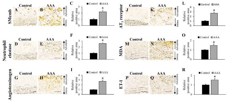
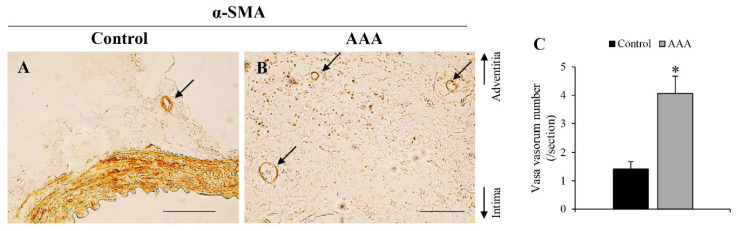
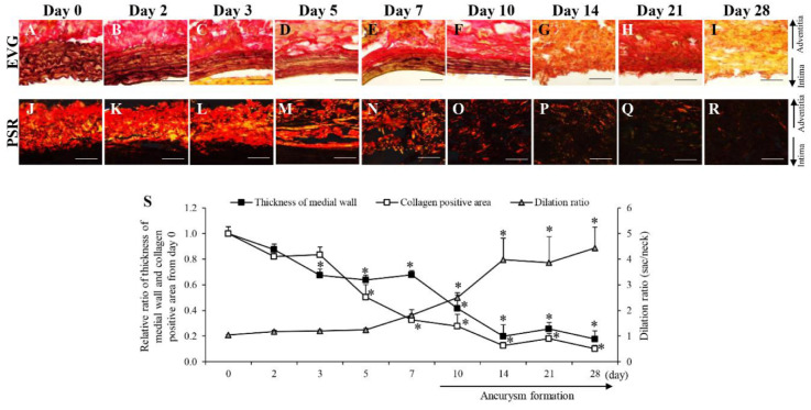
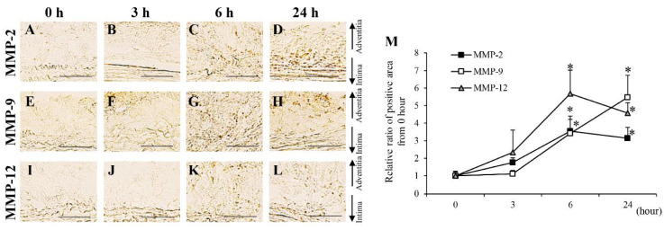
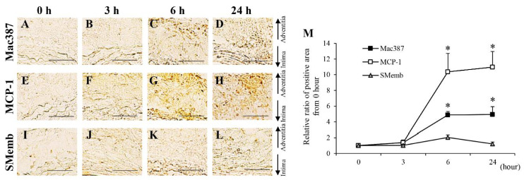
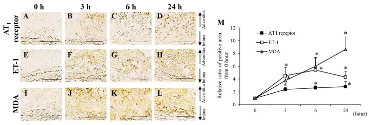
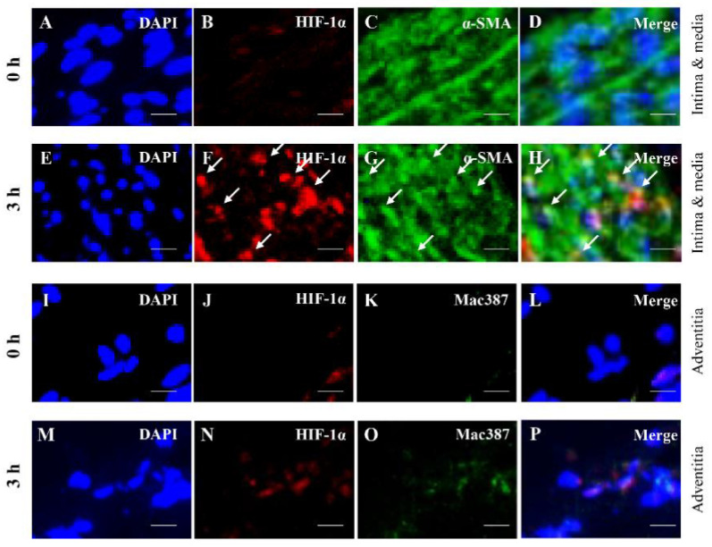
Hypoperfusion due to vasa vasorum stenosis can cause wall hypoxia and abdominal aortic aneurysm (AAA) development. Even though hypoperfusion is an important contributor toward pathological changes in AAA, the correlation between hypoperfusion and AAA is not fully understood. In this study, a time-dependent semi-quantitative pathological analysis of hypoperfusion-induced aortic wall changes was performed to understand the mechanisms underlying the gradual degradation of the aortic wall leading to AAA formation. AAA-related factors evaluated in this study were grouped according to the timing of dynamic change, and five groups were formed as follows: first group: angiotensin II type 1 receptor, endothelin-1 (ET-1), and malondialdehyde (MDA); second group: matrix metalloproteinase (MMP)-2, -9, -12, M1 macrophages (Mac387+ cells), and monocyte chemotactic protein-1; third group: synthetic smooth muscle cells (SMCs); fourth group: neutrophil elastase, contractile SMCs, and angiotensinogen; and the fifth group: M2 macrophages (CD163+ cells). Hypoxia-inducible factor-1α, ET-1, MDA, and MMP-9 were colocalized with alpha-smooth muscle actin cells in 3 h, suggesting that hypoperfusion-induced hypoxia directly affects the activities of contractile SMCs in the initial stage of AAA. Time-dependent pathological analysis clarified the cascade of AAA-related factors. These findings provide clues for understanding complicated multistage pathologies in AAA.
血管滋养管狭窄导致的灌注不足可引起血管壁缺氧和腹主动脉瘤(AAA)的发生。尽管灌注不足是AAA病理变化的一个重要因素,但灌注不足与AAA之间的相关性尚未完全明确。在本研究中,进行了灌注不足诱导的主动脉壁变化的时间依赖性半定量病理分析,以了解导致AAA形成的主动脉壁逐渐退化的潜在机制。本研究中评估的与AAA相关的因素根据动态变化的时间进行分组,共形成五组如下:第一组:血管紧张素II 1型受体、内皮素-1(ET-1)和丙二醛(MDA);第二组:基质金属蛋白酶(MMP)-2、-9、-12、M1巨噬细胞(Mac387+细胞)和单核细胞趋化蛋白-1;第三组:合成型平滑肌细胞(SMC);第四组:中性粒细胞弹性蛋白酶、收缩型SMC和血管紧张素原;第五组:M2巨噬细胞(CD163+细胞)。缺氧诱导因子-1α、ET-1、MDA和MMP-9在3小时内与α-平滑肌肌动蛋白细胞共定位,表明灌注不足诱导的缺氧在AAA初始阶段直接影响收缩型SMC的活性。时间依赖性病理分析阐明了与AAA相关因素的级联反应。这些发现为理解AAA复杂的多阶段病理提供了线索。