Universitätsmedizin Greifswald, Klinik und Poliklinik für Neurochirurgie, Sauerbruchstraße, 17475, Greifswald, Germany.
Acta Neurochir (Wien). 2021 Jun;163(6):1577-1581. doi: 10.1007/s00701-021-04788-1. Epub 2021 Mar 5.
Instrumentation of the lumbosacral region is one of the more challenging regions due to the complex anatomical structures and biomechanical forces. Screw insertion can be done both navigated and based on X-ray verification. In this study, we demonstrate a fast and reliable open, low exposure X-ray-guided technique of iliac screw placement.
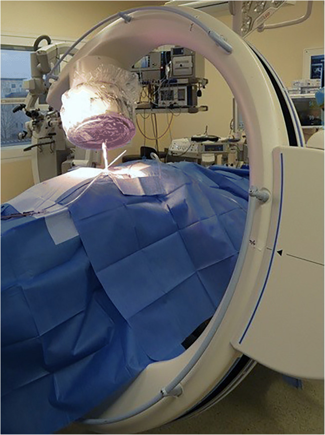
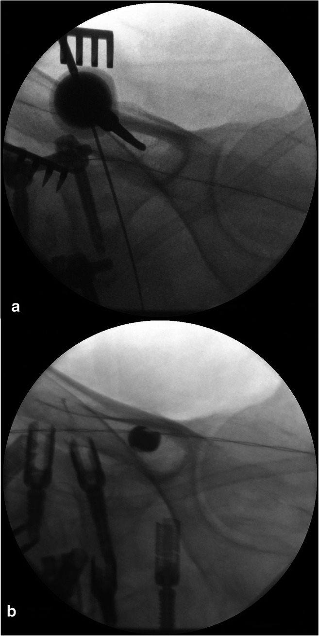
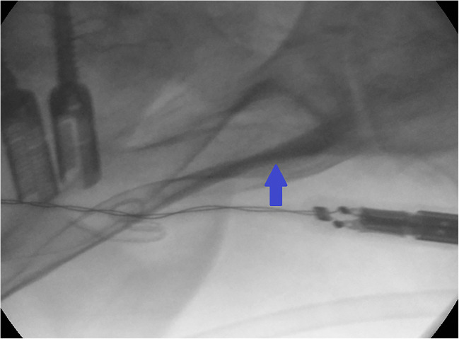
Between October 2016 and August 2019, 48 patients underwent sacropelvic fixation in tear-drop technique. Screw insertion was performed in open technique by using an X-ray converter angulated 25-30° in coronal and sagittal view. The anatomical insertion point was the posterior superior iliac spine. Verification of correct screw placement was done by intraoperative 3D scan.
In total, 95 iliac screws were placed in tear-drop technique with a correct placement in 98.1%.
The tear-drop technique showed a proper screw position in the intraoperative 3D scan and therefore may be considered an alternative technique to the navigated screw placement.
腰骶部是一个更具挑战性的区域,因为其具有复杂的解剖结构和生物力学。螺钉插入可以通过导航和 X 射线验证来完成。在这项研究中,我们展示了一种快速可靠的开放、低暴露 X 射线引导的髂骨螺钉放置技术。
2016 年 10 月至 2019 年 8 月,48 例患者采用泪滴技术行骨盆骶骨固定术。通过使用冠状位和矢状位成角 25-30°的 X 射线转换器,在开放技术中进行螺钉插入。解剖插入点为后上髂棘。通过术中 3D 扫描验证正确的螺钉放置。
共采用泪滴技术放置 95 枚髂骨螺钉,术中 3D 扫描显示螺钉位置正确者占 98.1%。
泪滴技术在术中 3D 扫描中显示出适当的螺钉位置,因此可以被认为是一种替代导航螺钉放置的技术。