Department of Orthopaedic Surgery, Graduate School of Medical Science, Kanazawa University, 13-1 Takaramachi, Kanazawa, 920-0934, Japan.
Department of Orthopaedic Surgery, National Hospital Organization, Kanazawa Medical Center, Kanazawa, Japan.
J Int Soc Sports Nutr. 2021 Mar 6;18(1):21. doi: 10.1186/s12970-021-00418-4.
Electromyography (EMG) has been used for evaluating skeletal muscle activity during pitching. However, it is difficult to observe the influence of movement on skeletal muscle activity in deep-lying regions of the trunk and extremities using EMG. An alternative method that may be used is the measurement of glucose metabolism of skeletal muscle using positron emission tomography-computed tomography (PET-CT). This technique is a reliable measure of muscle metabolism, demonstrating a high correlation with the intensity of muscle activity. This study aimed to evaluate whole-body skeletal muscle metabolism during pitching using PET-CT.
Ten uninjured, skilled, adult pitchers, who were active at college or professional level, threw 40 baseballs at maximal effort before an intravenous injection of 37 MBq of F-fluorodeoxyglucose (FDG). Subsequently, additional 40 balls were pitched. PET-CT images were obtained 50 min after FDG injection, and regions of interest were defined within 72 muscles. The standardized uptake value (SUV) of FDG by muscle tissue per unit volume was calculated, and the mean SUV of the pitchers was compared with that of a healthy adult control group who did not exercise before the measurements. Statistical analysis was performed using a t-test, and P < 0.05 was considered statistically significant.
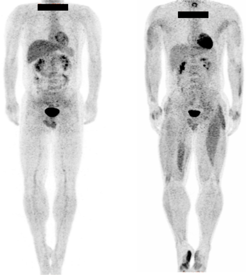
Whole-body PET images showed a significant increase in glucose metabolism in the muscle groups of the fingers and toes in both the throwing and non-throwing sides. Additionally, asymmetric increases in glucose metabolism were observed in the muscles of the thigh.
This is the first study to evaluate whole-body muscle metabolism during pitching using PET-CT. Our findings would be useful in determining the training required for pitchers, and can be further applied to other sporting activities that involve throwing.
肌电图(EMG)已被用于评估投球过程中的骨骼肌活动。然而,使用 EMG 很难观察到运动对躯干和四肢深部骨骼肌活动的影响。另一种可能的方法是使用正电子发射断层扫描-计算机断层扫描(PET-CT)测量骨骼肌的葡萄糖代谢。该技术是肌肉代谢的可靠测量方法,与肌肉活动强度具有高度相关性。本研究旨在使用 PET-CT 评估投球过程中的全身骨骼肌代谢。
10 名未受伤、技术熟练、处于大学或职业水平的成年投手,在静脉注射 37MBq F-氟脱氧葡萄糖(FDG)前,最大努力投掷 40 个棒球。随后,再投掷 40 个球。FDG 注射后 50 分钟获得 PET-CT 图像,并在 72 块肌肉内定义感兴趣区。计算肌肉组织单位体积内 FDG 的标准化摄取值(SUV),并将投手的平均 SUV 与未进行测量前的健康成年对照组进行比较。使用 t 检验进行统计分析,P<0.05 被认为具有统计学意义。
全身 PET 图像显示,投掷侧和非投掷侧的手指和脚趾肌肉群的葡萄糖代谢明显增加。此外,大腿肌肉也观察到葡萄糖代谢的不对称增加。
这是首次使用 PET-CT 评估投球过程中的全身肌肉代谢。我们的研究结果有助于确定投手所需的训练,并且可以进一步应用于其他涉及投掷的运动项目。