Zhang Rui, Xu Tao, Xia Yu, Wang Zhi, Li Xingrui, Chen Wen
Department of Thyroid and Breast Surgery, Wuhan No.1 Hospital, Wuhan, China.
Department of Thyroid and Breast Surgery, Tongji Hospital, Tongji Medical College, Huazhong University of Science and Technology, Wuhan, China.
Front Oncol. 2021 Feb 12;10:581733. doi: 10.3389/fonc.2020.581733. eCollection 2020.
High expression of integral membrane protein 2A (ITM2A) was reported to be associated with favorable prognosis in several solid tumors including breast cancer. This study aimed to investigate the role of ITM2A in breast cancer, especially in respect to tumor microenvironment.
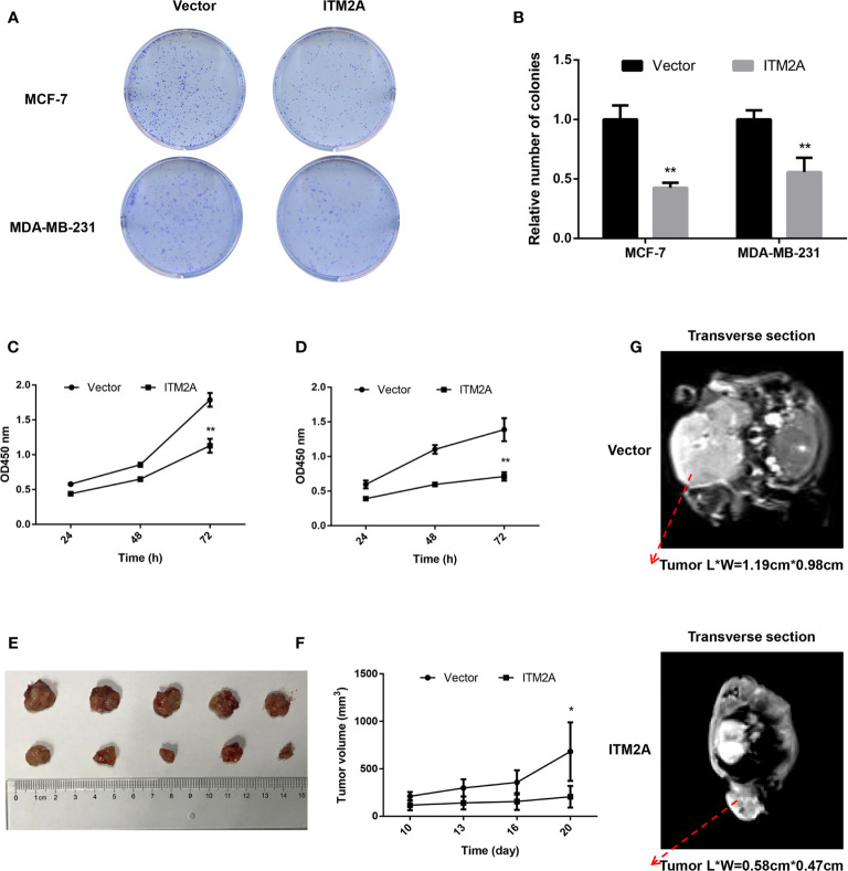
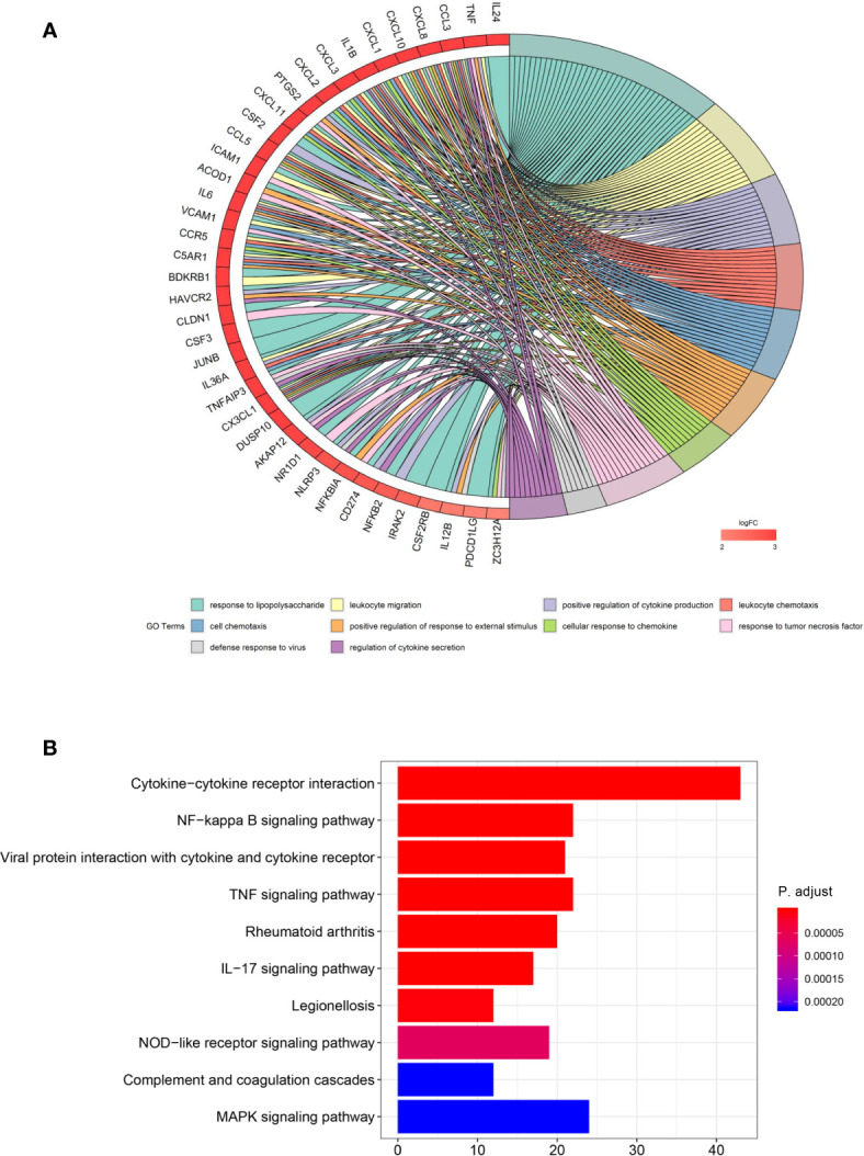
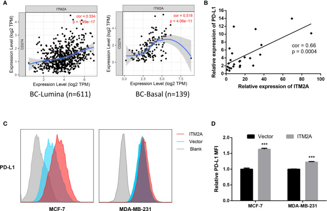
ITM2A expression was evaluated based on qRT-PCR results on breast cancer specimens, as well as TCGA and GEO datasets. The influence of ITM2A expression on breast cancer cell proliferation and tumor growth were evaluated by CCK-8 assay, clonogenic assay, and murine xenograft models. Transwell assay was performed to observe the changes of invasion and migration capacity in breast cancer cells. To determine the biological functions of ITM2A, differentially expressed genes (DEGs) were screened based on RNA-sequencing data of MCF-7 cells overexpressed ITM2A. Then, functional annotation on DEGs was given by Gene Ontology and KEGG analysis. The stimulation on programmed cell death ligand 1 (PD-L1) expression when ITM2A overexpressed was determined by flow cytometry. Meanwhile, the correlation on expression levels between PD-L1 and ITM2A was tested qRT-PCR on 24 breast cancer tissues, as well as public database.
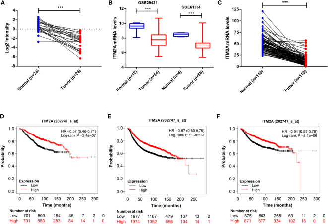
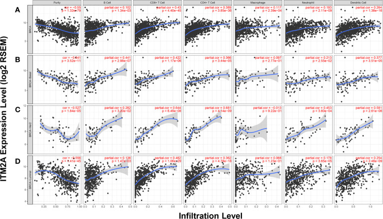
We demonstrated that ITM2A was frequently downregulated in breast cancer. Patients with high expression levels of ITM2A had longer overall survival and relapse free survival. Overexpression of ITM2A inhibited proliferation and impaired cells capacity of invasion and migration and . The DEGs in breast cancer cells overexpressed ITM2A were found to be associated with immunity responses. Moreover, ITM2A was found to facilitate breast cancer cells to express PD-L1. The correlation between PD-L1 and ITM2A was verified with both qRT-PCR assay and public database. Additionally, it was found that breast cancer had higher ITM2A expression frequently had more tumor-infiltrating lymphocytes (TILs).
In summary, we found that high expression of ITM2A reduced the aggressivity of breast cancer cells and had a favorable effect on outcomes of patients with breast cancer. Moreover, ITM2A induced PD-L1 expression in breast cancer cells was accompanied with higher TILs numbers in tumor microenvironment.
据报道,整合膜蛋白2A(ITM2A)的高表达与包括乳腺癌在内的几种实体瘤的良好预后相关。本研究旨在探讨ITM2A在乳腺癌中的作用,特别是在肿瘤微环境方面。
基于乳腺癌标本以及TCGA和GEO数据集的qRT-PCR结果评估ITM2A表达。通过CCK-8测定、克隆形成测定和小鼠异种移植模型评估ITM2A表达对乳腺癌细胞增殖和肿瘤生长的影响。进行Transwell测定以观察乳腺癌细胞侵袭和迁移能力的变化。为了确定ITM2A的生物学功能,基于过表达ITM2A的MCF-7细胞的RNA测序数据筛选差异表达基因(DEG)。然后,通过基因本体论和KEGG分析对DEG进行功能注释。通过流式细胞术确定ITM2A过表达时对程序性细胞死亡配体1(PD-L1)表达的刺激。同时,通过对24个乳腺癌组织以及公共数据库进行qRT-PCR检测PD-L1和ITM2A表达水平之间的相关性。
我们证明ITM2A在乳腺癌中经常下调。ITM2A表达水平高的患者总生存期和无复发生存期更长。ITM2A的过表达抑制增殖并损害细胞的侵袭和迁移能力。发现过表达ITM2A的乳腺癌细胞中的DEG与免疫反应相关。此外,发现ITM2A促进乳腺癌细胞表达PD-L1。通过qRT-PCR测定和公共数据库验证了PD-L1和ITM2A之间的相关性。此外,发现ITM2A表达较高的乳腺癌通常具有更多的肿瘤浸润淋巴细胞(TIL)。
总之,我们发现ITM2A的高表达降低了乳腺癌细胞的侵袭性,并对乳腺癌患者的预后产生了有利影响。此外,ITM2A诱导乳腺癌细胞中PD-L1表达伴随着肿瘤微环境中更高的TIL数量。