Ma Guofo, Kang Jie, Qiao Ning, Zhang Bochao, Chen Xuzhu, Li Guilin, Gao Zhixian, Gui Songbai
Department of Neurosurgery, Beijing Tiantan Hospital, Capital Medical University, Beijing, China.
Department of Radiology, Beijing Tiantan Hospital, Capital Medical University, Beijing, China.
Front Oncol. 2021 Feb 17;10:599888. doi: 10.3389/fonc.2020.599888. eCollection 2020.
Craniopharyngiomas (CPs) are benign tumors, complete tumor resection is considered to be the optimal treatment. However, although histologically benign, the local invasiveness of CPs commonly contributes to incomplete resection and a poor prognosis. At present, some advocate less aggressive surgery combined with radiotherapy as a more reasonable and effective means of protecting hypothalamus function and preventing recurrence in patients with tight tumor adhesion to the hypothalamus. Hence, if a method can be developed to predict the invasiveness of CP preoperatively, it will help in the development of a more personalized surgical strategy. The aim of the study was to report a radiomics-clinical nomogram for the individualized preoperative prediction of the invasiveness of adamantinomatous CP (ACPs) before surgery.
In total, 1,874 radiomics features were extracted from whole tumors on contrast-enhanced T1-weighted images. A support vector machine trained a predictive model that was validated using receiver operating characteristic (ROC) analysis on an independent test set. Moreover, a nomogram was constructed incorporating clinical characteristics and the radiomics signature for individual prediction.
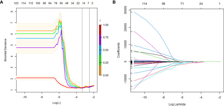
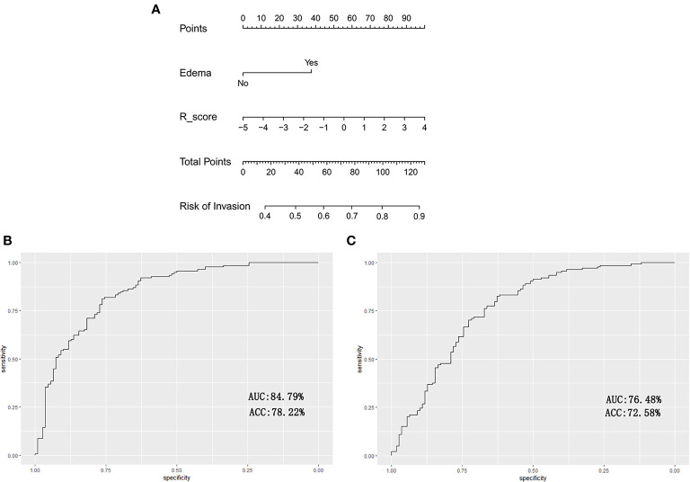
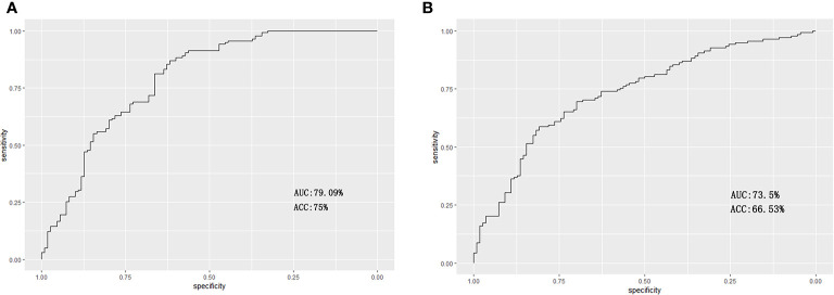
Eleven features associated with the invasiveness of ACPs were selected by using the least absolute shrinkage and selection operator (LASSO) method. These features yielded area under the curve (AUC) values of 79.09 and 73.5% for the training and test sets, respectively. The nomogram incorporating peritumoral edema and the radiomics signature yielded good calibration in the training and test sets with the AUCs of 84.79 and 76.48%, respectively.
The developed model yields good performance, indicating that the invasiveness of APCs can be predicted using noninvasive radiological data. This reliable, noninvasive tool can help clinical decision making and improve patient prognosis.
颅咽管瘤(CPs)是良性肿瘤,完整切除肿瘤被认为是最佳治疗方法。然而,尽管CPs在组织学上是良性的,但其局部侵袭性通常导致切除不完全和预后不良。目前,一些人主张采用较保守的手术联合放疗,作为保护下丘脑功能和防止肿瘤与下丘脑紧密粘连患者复发的更合理有效的方法。因此,如果能开发出一种术前预测CPs侵袭性的方法,将有助于制定更个性化的手术策略。本研究的目的是报告一种用于术前个体化预测造釉细胞瘤型颅咽管瘤(ACPs)侵袭性的影像组学-临床列线图。
总共从增强T1加权图像上的全肿瘤中提取了1874个影像组学特征。使用支持向量机训练预测模型,并在独立测试集上通过受试者操作特征(ROC)分析进行验证。此外,构建了一个列线图,纳入临床特征和影像组学特征进行个体预测。
使用最小绝对收缩和选择算子(LASSO)方法选择了11个与ACPs侵袭性相关的特征。这些特征在训练集和测试集上的曲线下面积(AUC)值分别为79.09%和73.5%。纳入瘤周水肿和影像组学特征的列线图在训练集和测试集上具有良好的校准性,AUC分别为84.79%和76.48%。
所开发的模型性能良好,表明可以使用无创放射学数据预测APCs的侵袭性。这种可靠的无创工具可以帮助临床决策并改善患者预后。