• 文献检索
  • 文档翻译
  • 深度研究
  • 学术资讯
  • Suppr Zotero 插件Zotero 插件
  • 邀请有礼
  • 套餐&价格
  • 历史记录
应用&插件
Suppr Zotero 插件Zotero 插件浏览器插件Mac 客户端Windows 客户端微信小程序
定价
高级版会员购买积分包购买API积分包
服务
文献检索文档翻译深度研究API 文档MCP 服务
关于我们
关于 Suppr公司介绍联系我们用户协议隐私条款
关注我们

Suppr 超能文献

核心技术专利:CN118964589B侵权必究
粤ICP备2023148730 号-1Suppr @ 2026

文献检索

告别复杂PubMed语法,用中文像聊天一样搜索,搜遍4000万医学文献。AI智能推荐,让科研检索更轻松。

立即免费搜索

文件翻译

保留排版,准确专业,支持PDF/Word/PPT等文件格式,支持 12+语言互译。

免费翻译文档

深度研究

AI帮你快速写综述,25分钟生成高质量综述,智能提取关键信息,辅助科研写作。

立即免费体验

基于磁共振图像的影像组学方法预测垂体腺瘤对海绵窦的侵袭

Preoperative prediction of cavernous sinus invasion by pituitary adenomas using a radiomics method based on magnetic resonance images.

机构信息

Neurosurgery, Beijing Tiantan Hospital, Capital Medical University, Beijing, 100050, China.

CAS Key Laboratory of Molecular Imaging, Institute of Automation, Beijing, 100190, China.

出版信息

Eur Radiol. 2019 Mar;29(3):1625-1634. doi: 10.1007/s00330-018-5725-3. Epub 2018 Sep 25.

DOI:10.1007/s00330-018-5725-3
PMID:30255254
原文链接:https://pmc.ncbi.nlm.nih.gov/articles/PMC6510860/
Abstract

OBJECTIVES

To predict cavernous sinus (CS) invasion by pituitary adenomas (PAs) pre-operatively using a radiomics method based on contrast-enhanced T1 (CE-T1) and T2-weighted magnetic resonance (MR) imaging.

METHODS

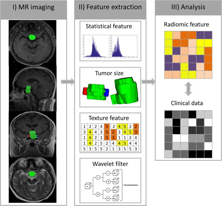
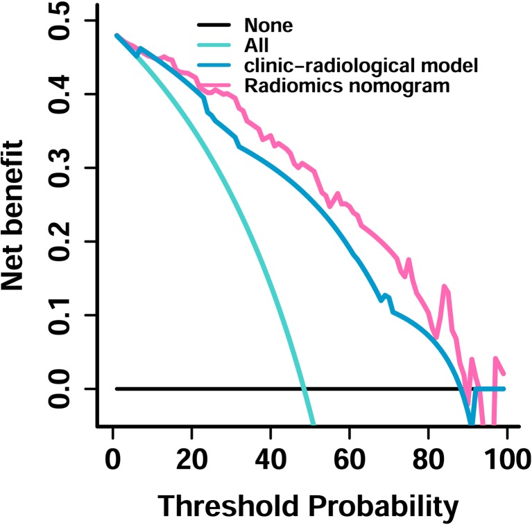
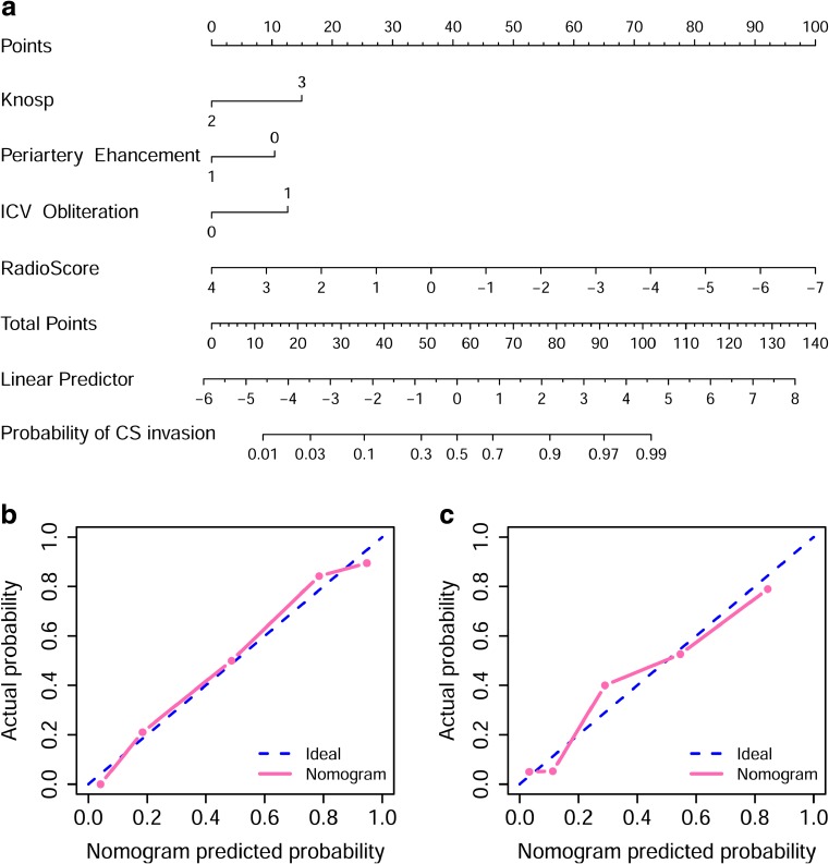
A total of 194 patients with Knosp grade two and three PAs (training set: n = 97; test set: n = 97) were enrolled in this retrospective study. From CE-T1 and T2 MR images, 2553 quantitative imaging features were extracted. To select the most informative features, least absolute shrinkage and selection operator (LASSO) was performed. Subsequently, a linear support vector machine (SVM) was used to fit the predictive model. Furthermore, a nomogram was constructed by incorporating clinico-radiological risk factors and radiomics signature, and the clinical usefulness of the nomogram was validated using decision curve analysis (DCA).

RESULTS

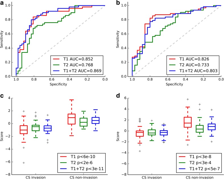
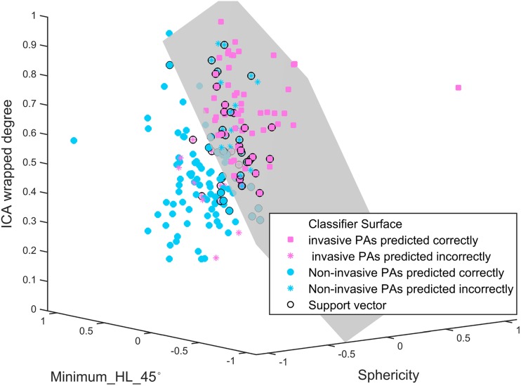
Three imaging features were selected in the training set, based on which the radiomics model yielded area under the curve (AUC) values of 0.852 and 0.826 for the training and test sets. The nomogram based on the radiomics signature and the clinico-radiological risk factors yielded an AUC of 0.899 in the training set and 0.871 in the test set.

CONCLUSIONS

The nomogram developed in this study might aid neurosurgeons in the pre-operative prediction of CS invasion by Knosp grade two and three PAs, which might contribute to creating surgical strategies.

KEY POINTS

• Pre-operative diagnosis of CS invasion by PAs might affect creating surgical strategies • MRI might help for diagnosis of CS invasion by PAs before surgery • Radiomics might improve the CS invasion detection by MR images.

摘要

目的

利用基于对比增强 T1(CE-T1)和 T2 加权磁共振(MR)成像的放射组学方法,术前预测垂体腺瘤(PA)的海绵窦(CS)侵袭。

方法

本回顾性研究共纳入 194 例 Knosp 分级为 2 级和 3 级的 PA 患者(训练集:n=97;测试集:n=97)。从 CE-T1 和 T2 MR 图像中提取了 2553 个定量成像特征。为了选择最具信息量的特征,进行了最小绝对收缩和选择算子(LASSO)。随后,使用线性支持向量机(SVM)拟合预测模型。此外,通过纳入临床-放射学风险因素和放射组学特征构建了列线图,并通过决策曲线分析(DCA)验证了该列线图的临床实用性。

结果

在训练集中,基于三个影像学特征,放射组学模型在训练集和测试集的 AUC 值分别为 0.852 和 0.826。基于放射组学特征和临床-放射学风险因素的列线图在训练集的 AUC 值为 0.899,在测试集的 AUC 值为 0.871。

结论

本研究中开发的列线图可能有助于神经外科医生术前预测 Knosp 分级为 2 级和 3 级的 PA 对 CS 的侵袭,这可能有助于制定手术策略。

关键点

• PA 对 CS 的侵袭术前诊断可能会影响制定手术策略。

• MRI 可能有助于手术前诊断 PA 对 CS 的侵袭。

• 放射组学可能会提高 MR 图像对 CS 侵袭的检测。

https://cdn.ncbi.nlm.nih.gov/pmc/blobs/cb70/6510860/3a1c76b6819a/330_2018_5725_Fig5_HTML.jpg
https://cdn.ncbi.nlm.nih.gov/pmc/blobs/cb70/6510860/241cb6b5704d/330_2018_5725_Fig1_HTML.jpg
https://cdn.ncbi.nlm.nih.gov/pmc/blobs/cb70/6510860/c31a1a827b5a/330_2018_5725_Fig2_HTML.jpg
https://cdn.ncbi.nlm.nih.gov/pmc/blobs/cb70/6510860/e43ef6e86c5b/330_2018_5725_Fig3_HTML.jpg
https://cdn.ncbi.nlm.nih.gov/pmc/blobs/cb70/6510860/2119f46711aa/330_2018_5725_Fig4_HTML.jpg
https://cdn.ncbi.nlm.nih.gov/pmc/blobs/cb70/6510860/3a1c76b6819a/330_2018_5725_Fig5_HTML.jpg

相似文献

1
Preoperative prediction of cavernous sinus invasion by pituitary adenomas using a radiomics method based on magnetic resonance images.基于磁共振图像的影像组学方法预测垂体腺瘤对海绵窦的侵袭
Eur Radiol. 2019 Mar;29(3):1625-1634. doi: 10.1007/s00330-018-5725-3. Epub 2018 Sep 25.
2
Non-invasive radiomics approach potentially predicts non-functioning pituitary adenomas subtypes before surgery.非侵入性放射组学方法可能在手术前预测无功能垂体腺瘤亚型。
Eur Radiol. 2018 Sep;28(9):3692-3701. doi: 10.1007/s00330-017-5180-6. Epub 2018 Mar 23.
3
A radiomics nomogram may improve the prediction of IDH genotype for astrocytoma before surgery.一个放射组学列线图可能会提高术前预测星形细胞瘤 IDH 基因型的能力。
Eur Radiol. 2019 Jul;29(7):3325-3337. doi: 10.1007/s00330-019-06056-4. Epub 2019 Apr 10.
4
Radiomics analysis of multiparametric MRI for prediction of pathological complete response to neoadjuvant chemoradiotherapy in locally advanced rectal cancer.多参数 MRI 的放射组学分析预测局部晚期直肠癌新辅助放化疗的病理完全缓解。
Eur Radiol. 2019 Mar;29(3):1211-1220. doi: 10.1007/s00330-018-5683-9. Epub 2018 Aug 20.
5
Development of a Nomogram Based on Preoperative Bi-Parametric MRI and Blood Indices for the Differentiation Between Cystic-Solid Pituitary Adenoma and Craniopharyngioma.基于术前双参数磁共振成像和血液指标的列线图在囊性实性垂体腺瘤与颅咽管瘤鉴别诊断中的应用
Front Oncol. 2021 Jul 9;11:709321. doi: 10.3389/fonc.2021.709321. eCollection 2021.
6
Pre-operative MRI predictors of hormonal remission status post pituitary adenoma resection.术前 MRI 预测垂体腺瘤切除术后激素缓解状态。
Clin Imaging. 2019 May-Jun;55:29-34. doi: 10.1016/j.clinimag.2019.01.020. Epub 2019 Jan 25.
7
A machine learning model to precisely immunohistochemically classify pituitary adenoma subtypes with radiomics based on preoperative magnetic resonance imaging.基于术前磁共振成像的放射组学机器学习模型对垂体腺瘤亚型进行精确免疫组化学分类。
Eur J Radiol. 2020 Apr;125:108892. doi: 10.1016/j.ejrad.2020.108892. Epub 2020 Feb 13.
8
Comparative analysis of intraoperative and imaging features of invasive growth in pituitary adenomas.侵袭性垂体腺瘤术中与影像学特征的对比分析。
Eur J Endocrinol. 2024 Jun 5;190(6):489-500. doi: 10.1093/ejendo/lvae059.
9
Development and validation of an MRI-based radiomic signature for the preoperative prediction of treatment response in patients with invasive functional pituitary adenoma.基于 MRI 的放射组学特征的建立与验证:术前预测侵袭性功能性垂体腺瘤患者的治疗反应。
Eur J Radiol. 2019 Dec;121:108647. doi: 10.1016/j.ejrad.2019.108647. Epub 2019 Sep 7.
10
Radiomics analysis allows for precise prediction of silent corticotroph adenoma among non-functioning pituitary adenomas.影像组学分析可精准预测无功能垂体腺瘤中的静默促肾上腺皮质激素腺瘤。
Eur Radiol. 2022 Mar;32(3):1570-1578. doi: 10.1007/s00330-021-08361-3. Epub 2021 Nov 27.

引用本文的文献

1
MRI-based interpretable clinicoradiological and radiomics machine learning model for preoperative prediction of pituitary macroadenomas consistency: a dual-center study.基于MRI的可解释性临床放射学和放射组学机器学习模型用于垂体大腺瘤一致性的术前预测:一项双中心研究。
Neuroradiology. 2025 Jul 9. doi: 10.1007/s00234-025-03698-8.
2
Preoperatively Predicting PIT1 Expression in Pituitary Adenomas Using Habitat, Intra-tumoral and Peri-tumoral Radiomics Based on MRI.基于MRI,利用瘤体、瘤内及瘤周影像组学特征术前预测垂体腺瘤中的PIT1表达
J Imaging Inform Med. 2025 Feb 4. doi: 10.1007/s10278-024-01376-4.
3
Preoperative prediction of pituitary neuroendocrine tumor invasion using multiparametric MRI radiomics.

本文引用的文献

1
Radiomics strategies for risk assessment of tumour failure in head-and-neck cancer.头颈部肿瘤肿瘤失败风险评估的放射组学策略。
Sci Rep. 2017 Aug 31;7(1):10117. doi: 10.1038/s41598-017-10371-5.
2
Different Volumetric Measurement Methods for Pituitary Adenomas and Their Crucial Clinical Significance.不同的垂体腺瘤体积测量方法及其关键的临床意义。
Sci Rep. 2017 Jan 18;7:40792. doi: 10.1038/srep40792.
3
MR Imaging Radiomics Signatures for Predicting the Risk of Breast Cancer Recurrence as Given by Research Versions of MammaPrint, Oncotype DX, and PAM50 Gene Assays.
使用多参数MRI影像组学对垂体神经内分泌肿瘤侵袭进行术前预测
Front Oncol. 2025 Jan 9;14:1475950. doi: 10.3389/fonc.2024.1475950. eCollection 2024.
4
Peritumoral vessel characteristics on magnetic resonance venography reflect the sinus invasion status of para-sinus meningioma.磁共振静脉血管造影上的瘤周血管特征反映了鼻窦旁脑膜瘤的窦侵犯状态。
Quant Imaging Med Surg. 2024 Dec 5;14(12):8183-8195. doi: 10.21037/qims-24-278. Epub 2024 Oct 21.
5
Applications and Integration of Radiomics for Skull Base Oncology.颅底肿瘤放射组学的应用与整合。
Adv Exp Med Biol. 2024;1462:285-305. doi: 10.1007/978-3-031-64892-2_17.
6
The current state of MRI-based radiomics in pituitary adenoma: promising but challenging.基于 MRI 的脑垂体瘤放射组学的现状:前景广阔但极具挑战。
Front Endocrinol (Lausanne). 2024 Sep 20;15:1426781. doi: 10.3389/fendo.2024.1426781. eCollection 2024.
7
Diagnosis of choroid plexus papilloma: Current perspectives and future directions.脉络丛乳头状瘤的诊断:当前观点与未来方向
Cancer Pathog Ther. 2023 Sep 26;2(3):173-179. doi: 10.1016/j.cpt.2023.09.005. eCollection 2024 Jul.
8
Radiomics of pituitary adenoma using computer vision: a review.基于计算机视觉的垂体腺瘤影像组学研究:综述
Med Biol Eng Comput. 2024 Dec;62(12):3581-3597. doi: 10.1007/s11517-024-03163-3. Epub 2024 Jul 16.
9
The Applications of Artificial Intelligence in Digestive System Neoplasms: A Review.人工智能在消化系统肿瘤中的应用:综述
Health Data Sci. 2023 Feb 6;3:0005. doi: 10.34133/hds.0005. eCollection 2023.
10
Predicting delayed remission in Cushing's disease using radiomics models: a multi-center study.使用影像组学模型预测库欣病的延迟缓解:一项多中心研究。
Front Oncol. 2024 Jan 9;13:1218897. doi: 10.3389/fonc.2023.1218897. eCollection 2023.
用于预测MammaPrint、Oncotype DX和PAM50基因检测研究版本所给出的乳腺癌复发风险的磁共振成像放射组学特征
Radiology. 2016 Nov;281(2):382-391. doi: 10.1148/radiol.2016152110. Epub 2016 May 5.
4
Development and Validation of a Radiomics Nomogram for Preoperative Prediction of Lymph Node Metastasis in Colorectal Cancer.基于影像组学的直肠癌淋巴结转移术前预测列线图模型的建立与验证。
J Clin Oncol. 2016 Jun 20;34(18):2157-64. doi: 10.1200/JCO.2015.65.9128. Epub 2016 May 2.
5
Early-Stage Non-Small Cell Lung Cancer: Quantitative Imaging Characteristics of (18)F Fluorodeoxyglucose PET/CT Allow Prediction of Distant Metastasis.早期非小细胞肺癌:(18)F氟脱氧葡萄糖PET/CT的定量成像特征可预测远处转移。
Radiology. 2016 Oct;281(1):270-8. doi: 10.1148/radiol.2016151829. Epub 2016 Apr 5.
6
Identifying the association between contrast enhancement pattern, surgical resection, and prognosis in anaplastic glioma patients.确定间变性胶质瘤患者的对比增强模式、手术切除与预后之间的关联。
Neuroradiology. 2016 Apr;58(4):367-74. doi: 10.1007/s00234-016-1640-y. Epub 2016 Jan 21.
7
Invasion of the cavernous sinus space in pituitary adenomas: endoscopic verification and its correlation with an MRI-based classification.垂体腺瘤侵袭海绵窦间隙:内镜验证及其与基于磁共振成像的分类的相关性
J Neurosurg. 2015 Apr;122(4):803-11. doi: 10.3171/2014.12.JNS141083. Epub 2015 Feb 6.
8
Decision curve analysis.决策曲线分析
JAMA. 2015 Jan 27;313(4):409-10. doi: 10.1001/jama.2015.37.
9
Anatomical localization of isocitrate dehydrogenase 1 mutation: a voxel-based radiographic study of 146 low-grade gliomas.异柠檬酸脱氢酶 1 突变的解剖定位:146 例低级别胶质瘤的基于体素的放射影像学研究。
Eur J Neurol. 2015 Feb;22(2):348-54. doi: 10.1111/ene.12578. Epub 2014 Oct 16.
10
Endoscopic endonasal transsellar approach for laterally extended pituitary adenomas: volumetric analysis of cavernous sinus invasion.经鼻内镜经蝶入路治疗向外侧扩展的垂体腺瘤:海绵窦侵犯的容积分析
Pituitary. 2015 Aug;18(4):518-24. doi: 10.1007/s11102-014-0604-7.