Department of Pulmonary Medicine, Rasoul Akram Hospital, Iran University of Medical Sciences, Tehran, Iran..
Department of Cardiology, Rasoul Akram Hospital, Iran University of Medical Sciences, Tehran, Iran.
Acta Biomed. 2021 Jan 28;92(1):e2021074. doi: 10.23750/abm.v92i1.9216.
Because of invasive nature of catheterization, using other noninvasive tools is more preferred to assess pulmonary arterial hypertension (PAH). The present study assessed the value of chest spiral CT scan and Doppler echocardiography compared to right heart catheterization (RHC) to predict PAH in patients with scleroderma.
This cross-sectional study was performed on 15 patients with limited scleroderma. All subjects underwent Doppler echocardiography (to assess PAP) and chest spiral CT scan without injection (to assess pulmonary trunk length or PUL), followed by RHC to assess PAH.
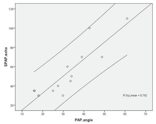
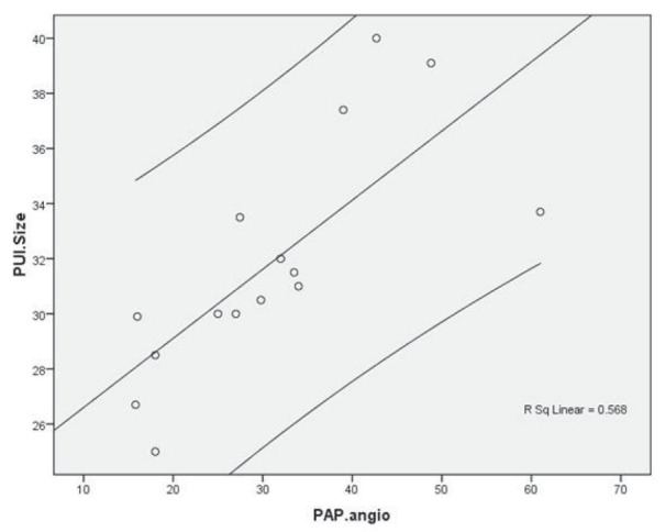
Comparing PUL in spiral CT scan with PAP in RHC yielded a sensitivity of 75.0% and a specificity of 100% for predicting PAH. Similarly, comparing PAP value in echocardiography with PAP in RHC achieved a sensitivity of 100% and a specificity of 63.6% to discriminate PAH from normal PAP condition. Analysis of the area under the ROC curve showed high power of CT scan to predict PAH (AUC = 1.000). The best cutoff point for PUL to predict PAH was 29.95 yielding a sensitivity of 100% and a specificity of 100%. Also, ROC curve analysis showed high value of echocardiography to discriminate PAH from normal PAP status (AUC = 0.841) that considering a cutoff value of 22.88 for PAP assessed by echocardiography reached to a sensitivity of 72.7% and a specificity of 100%. Conclusion: Both chest spiral CT scan and Doppler echocardiography are very useful to diagnose PAH and its severity in patients with scleroderma.
由于导管检查具有侵袭性,因此使用其他非侵入性工具来评估肺动脉高压(PAH)更为可取。本研究评估了胸部螺旋 CT 扫描和多普勒超声心动图与右心导管检查(RHC)相比,用于评估硬皮病患者 PAH 的价值。
这是一项横断面研究,共纳入 15 例局限性硬皮病患者。所有患者均接受多普勒超声心动图(评估肺动脉压[PAP])和胸部螺旋 CT 扫描(不注射造影剂,评估肺动脉干长度或 PUL),然后进行 RHC 以评估 PAH。
比较螺旋 CT 扫描中的 PUL 与 RHC 中的 PAP,预测 PAH 的敏感性为 75.0%,特异性为 100%。同样,比较超声心动图中的 PAP 值与 RHC 中的 PAP 值,鉴别 PAH 与正常 PAP 状态的敏感性为 100%,特异性为 63.6%。ROC 曲线下面积分析显示 CT 扫描预测 PAH 的效能较高(AUC=1.000)。预测 PAH 的最佳 PUL 截断值为 29.95,敏感性为 100%,特异性为 100%。ROC 曲线分析显示,超声心动图鉴别 PAH 与正常 PAP 状态的效能较高(AUC=0.841),考虑到超声心动图评估的 PAP 截断值为 22.88,敏感性为 72.7%,特异性为 100%。
胸部螺旋 CT 扫描和多普勒超声心动图均可非常有效地诊断硬皮病患者的 PAH 及其严重程度。