Department of Biological Sciences, Wright State University, Dayton, OH.
Odyssey Systems, Environmental Health Effects Laboratory, Navy Medical Research Unit, Dayton, Wright-Patterson Air Force Base, Dayton, OH.
J Gen Physiol. 2021 Apr 5;153(4). doi: 10.1085/jgp.202012637.
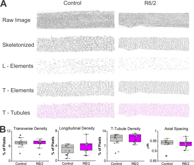
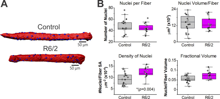
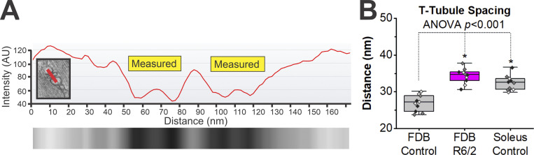
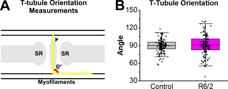
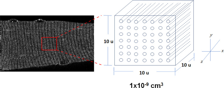
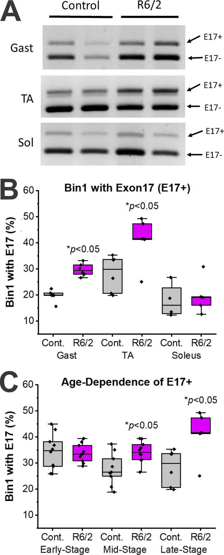
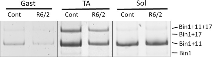
Huntington's disease (HD) is a fatal and progressive condition with severe debilitating motor defects and muscle weakness. Although classically recognized as a neurodegenerative disorder, there is increasing evidence of cell autonomous toxicity in skeletal muscle. We recently demonstrated that skeletal muscle fibers from the R6/2 model mouse of HD have a decrease in specific membrane capacitance, suggesting a loss of transverse tubule (t-tubule) membrane in R6/2 muscle. A previous report also indicated that Cav1.1 current was reduced in R6/2 skeletal muscle, suggesting defects in excitation-contraction (EC) coupling. Thus, we hypothesized that a loss and/or disruption of the skeletal muscle t-tubule system contributes to changes in EC coupling in R6/2 skeletal muscle. We used live-cell imaging with multiphoton confocal microscopy and transmission electron microscopy to assess the t-tubule architecture in late-stage R6/2 muscle and found no significant differences in the t-tubule system density, regularity, or integrity. However, electron microscopy images revealed that the cross-sectional area of t-tubules at the triad were 25% smaller in R6/2 compared with age-matched control skeletal muscle. Computer simulation revealed that the resulting decrease in the R6/2 t-tubule luminal conductance contributed to, but did not fully explain, the reduced R6/2 membrane capacitance. Analyses of bridging integrator-1 (Bin1), which plays a primary role in t-tubule formation, revealed decreased Bin1 protein levels and aberrant splicing of Bin1 mRNA in R6/2 muscle. Additionally, the distance between the t-tubule and sarcoplasmic reticulum was wider in R6/2 compared with control muscle, which was associated with a decrease in junctophilin 1 and 2 mRNA levels. Altogether, these findings can help explain dysregulated EC coupling and motor impairment in Huntington's disease.
亨廷顿病(HD)是一种致命且进行性的疾病,伴有严重的进行性运动缺陷和肌肉无力。尽管经典上被认为是一种神经退行性疾病,但越来越多的证据表明骨骼肌存在细胞自主毒性。我们最近证明,HD 的 R6/2 模型小鼠的骨骼肌纤维的特定膜电容降低,表明 R6/2 肌肉中的横管(t-管)膜丢失。先前的报告还表明,R6/2 骨骼肌中的 Cav1.1 电流减少,表明兴奋-收缩(EC)偶联缺陷。因此,我们假设骨骼肌 t-管系统的丢失和/或破坏导致 R6/2 骨骼肌 EC 偶联的变化。我们使用多光子共聚焦显微镜和透射电子显微镜进行活细胞成像,以评估晚期 R6/2 肌肉中的 t-管结构,发现 t-管系统的密度、规律性或完整性没有显着差异。然而,电子显微镜图像显示,与年龄匹配的对照骨骼肌相比,R6/2 中的三联体处 t-管的横截面积小 25%。计算机模拟表明,R6/2 t-管内腔电导率的降低导致但不能完全解释 R6/2 膜电容的降低。对主要参与 t-管形成的桥接整合素-1(Bin1)的分析表明,R6/2 肌肉中的 Bin1 蛋白水平降低和 Bin1 mRNA 的异常剪接。此外,与对照肌肉相比,R6/2 中的 t-管和肌浆网之间的距离更宽,这与 junctophilin 1 和 2 mRNA 水平的降低有关。总的来说,这些发现可以帮助解释亨廷顿病中失调的 EC 偶联和运动障碍。