Thayer School of Engineering at Dartmouth, Optics in Medicine, Hanover, New Hampshire, United States.
Geisel School of Medicine at Dartmouth, Department of Surgery, Hanover, New Hampshire, United States.
J Biomed Opt. 2019 Sep;24(9):1-12. doi: 10.1117/1.JBO.24.9.096003.
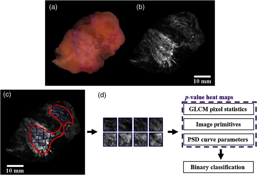
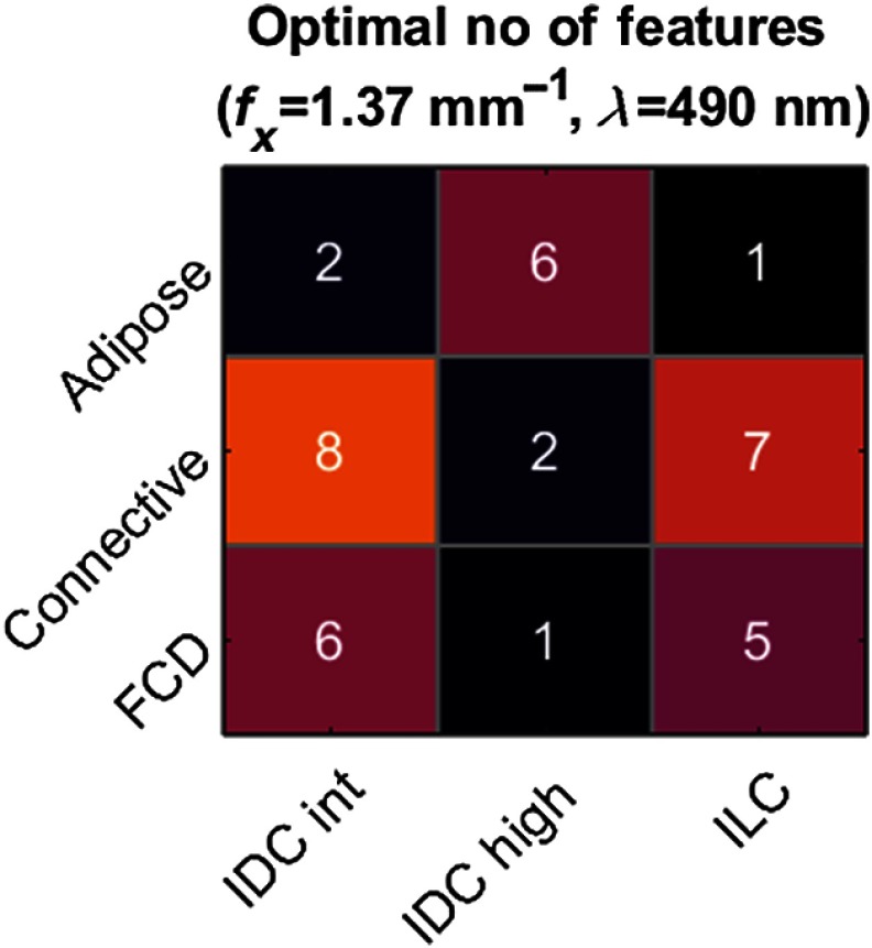
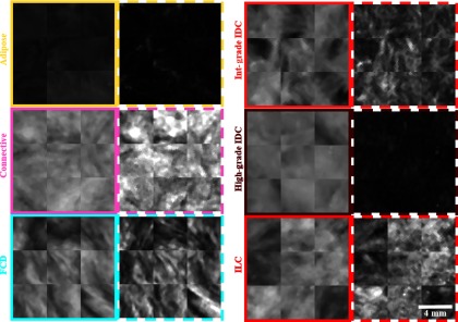
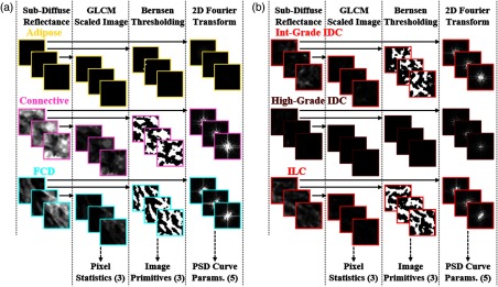
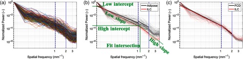
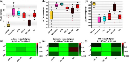
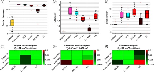
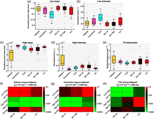
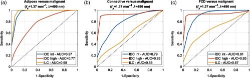
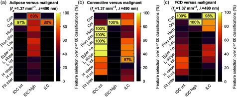
Subdiffuse spatial frequency domain imaging (sd-SFDI) data of 42 freshly excised, bread-loafed tumor resections from breast-conserving surgery (BCS) were evaluated using texture analysis and a machine learning framework for tissue classification. Resections contained 56 regions of interest (RoIs) determined by expert histopathological analysis. RoIs were coregistered with sd-SFDI data and sampled into ∼4 × 4 mm2 subimage samples of confirmed and homogeneous histological categories. Sd-SFDI reflectance textures were analyzed using gray-level co-occurrence matrix pixel statistics, image primitives, and power spectral density curve parameters. Texture metrics exhibited statistical significance (p-value < 0.05) between three benign and three malignant tissue subtypes. Pairs of benign and malignant subtypes underwent texture-based, binary classification with correlation-based feature selection. Classification performance was evaluated using fivefold cross-validation and feature grid searching. Classification using subdiffuse, monochromatic reflectance (illumination spatial frequency of fx = 1.37 mm − 1, optical wavelength of λ = 490 nm) achieved accuracies ranging from 0.55 (95% CI: 0.41 to 0.69) to 0.95 (95% CI: 0.90 to 1.00) depending on the benign–malignant diagnosis pair. Texture analysis of sd-SFDI data maintains the spatial context within images, is free of light transport model assumptions, and may provide an alternative, computationally efficient approach for wide field-of-view (cm2) BCS tumor margin assessment relative to pixel-based optical scatter or color properties alone.
利用纹理分析和机器学习框架对 42 个从保乳手术(BCS)新鲜切除的面包状肿瘤切除物的漫射空间频率域成像(sd-SFDI)数据进行评估,用于组织分类。切除物包含 56 个由专家组织病理学分析确定的感兴趣区域(ROI)。ROI 与 sd-SFDI 数据配准,并采样到经确认的同质组织学分类的约 4×4mm2 子图像样本中。使用灰度共生矩阵像素统计、图像基元和功率谱密度曲线参数分析 sd-SFDI 反射率纹理。纹理指标在三种良性和三种恶性组织亚型之间表现出统计学意义(p 值<0.05)。良性和恶性亚型对之间进行基于纹理的二进制分类,并进行基于相关性的特征选择。使用五重交叉验证和特征网格搜索评估分类性能。使用漫射、单色反射(照明空间频率 fx=1.37mm-1,光学波长 λ=490nm)的分类准确率范围为 0.55(95%CI:0.41 至 0.69)至 0.95(95%CI:0.90 至 1.00),具体取决于良性-恶性诊断对。sd-SFDI 数据的纹理分析保持了图像内的空间上下文,无需光传输模型假设,并且相对于单独的基于像素的光散射或颜色特性,可能为宽视场(cm2)BCS 肿瘤边界评估提供替代的、计算效率高的方法。