• 文献检索
  • 文档翻译
  • 深度研究
  • 学术资讯
  • Suppr Zotero 插件Zotero 插件
  • 邀请有礼
  • 套餐&价格
  • 历史记录
应用&插件
Suppr Zotero 插件Zotero 插件浏览器插件Mac 客户端Windows 客户端微信小程序
定价
高级版会员购买积分包购买API积分包
服务
文献检索文档翻译深度研究API 文档MCP 服务
关于我们
关于 Suppr公司介绍联系我们用户协议隐私条款
关注我们

Suppr 超能文献

核心技术专利:CN118964589B侵权必究
粤ICP备2023148730 号-1Suppr @ 2026

文献检索

告别复杂PubMed语法,用中文像聊天一样搜索,搜遍4000万医学文献。AI智能推荐,让科研检索更轻松。

立即免费搜索

文件翻译

保留排版,准确专业,支持PDF/Word/PPT等文件格式,支持 12+语言互译。

免费翻译文档

深度研究

AI帮你快速写综述,25分钟生成高质量综述,智能提取关键信息,辅助科研写作。

立即免费体验

中国大临床数据集乳腺癌患者中乳腺密度、年龄和乳腺病变类型之间的关系。

The relationship between breast density, age, and mammographic lesion type among Chinese breast cancer patients from a large clinical dataset.

机构信息

Department of Breast Imaging, Tianjin Medical University Cancer Institute and Hospital, Tianjin, China.

National Clinical Research Center for Cancer, Key Laboratory of Cancer Prevention and Therapy, Tianjin's Clinical Research Center for Cancer, Key Laboratory of Breast Cancer Prevention and Therapy, Tianjin Medical University, Ministry of Education, Tianjin, China.

出版信息

BMC Med Imaging. 2021 Mar 8;21(1):43. doi: 10.1186/s12880-021-00565-9.

DOI:10.1186/s12880-021-00565-9
PMID:33685388
原文链接:https://pmc.ncbi.nlm.nih.gov/articles/PMC7938487/
Abstract

BACKGROUND

The purpose of this study was to investigate the relationship between breast density, age, and mammographic lesion type among Chinese breast cancer patients included in a large clinical dataset.

METHODS

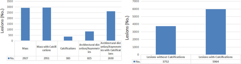
A review of mammographic images acquired between July 2014 and June 2017 from a total of 9716 retrospectively registered breast cancer patients was conducted. Mammographic breast density was defined according to the American College of Radiology Breast Imaging Reporting and Data System (ACR BI-RADS) 4-class density rating. Mammographic lesion types were defined according to the ACR BI-RADS, including mass, mass with calcifications, calcifications, architectural distortion/asymmetries, and architectural distortion/asymmetries with calcifications. Three experienced breast radiologists interpreted all mammograms. The chi-square (χ) test and Pearson correlation analyses were performed to assess the relationship between breast density, age, and mammographic lesion type.

RESULTS

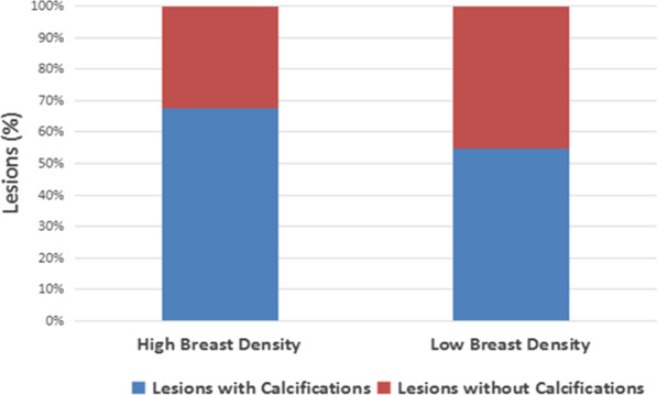
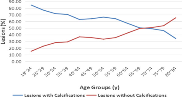
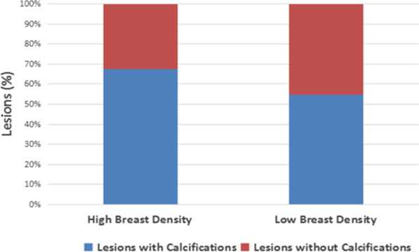
A significant inverse relationship was observed between the BI-RADS breast density rating given by radiologists and patient age (r = - 0.521, p < 0.01). The breast density distribution in breast cancer patients from China reversed at the age of 55 years, and exhibited one age peak in the age 55-59 year group. The percentage of lesions with calcifications decreased with increasing age (p < 0.01), and increased with increasing breast density (p < 0.01).

CONCLUSIONS

In general, we identified a relationship between patient breast density, age, and mammographic lesion type. This finding may provide a basis for clinical diagnoses and support development of breast cancer screening programs in China.

摘要

背景

本研究旨在探讨中国乳腺癌患者大临床数据集的乳腺密度、年龄与乳腺影像学病变类型之间的关系。

方法

回顾性分析 2014 年 7 月至 2017 年 6 月期间共 9716 例经注册的乳腺癌患者的乳腺 X 线摄影图像。乳腺密度根据美国放射学院乳腺影像学报告和数据系统(ACR BI-RADS)4 级密度分级标准定义。乳腺影像学病变类型根据 ACR BI-RADS 定义,包括肿块、肿块伴钙化、钙化、结构扭曲/不对称和结构扭曲/不对称伴钙化。由 3 名有经验的乳腺放射科医生对所有乳腺 X 线片进行解读。采用卡方检验(χ2 检验)和 Pearson 相关分析评估乳腺密度、年龄与乳腺影像学病变类型之间的关系。

结果

放射科医生给出的 BI-RADS 乳腺密度分级与患者年龄之间呈显著负相关(r = -0.521,p<0.01)。中国乳腺癌患者的乳腺密度分布在 55 岁时发生逆转,在 55-59 岁年龄组出现一个年龄高峰。伴有钙化的病变百分比随年龄的增加而降低(p<0.01),随乳腺密度的增加而增加(p<0.01)。

结论

总体而言,我们发现了患者的乳腺密度、年龄与乳腺影像学病变类型之间的关系。这一发现可能为临床诊断提供依据,并为中国的乳腺癌筛查计划提供支持。

https://cdn.ncbi.nlm.nih.gov/pmc/blobs/828f/7938487/8fc05946ac25/12880_2021_565_Fig6_HTML.jpg
https://cdn.ncbi.nlm.nih.gov/pmc/blobs/828f/7938487/73178c144b0f/12880_2021_565_Fig1_HTML.jpg
https://cdn.ncbi.nlm.nih.gov/pmc/blobs/828f/7938487/985039d0a34f/12880_2021_565_Fig2_HTML.jpg
https://cdn.ncbi.nlm.nih.gov/pmc/blobs/828f/7938487/ee756849e91e/12880_2021_565_Fig3_HTML.jpg
https://cdn.ncbi.nlm.nih.gov/pmc/blobs/828f/7938487/58964b56ac54/12880_2021_565_Fig4_HTML.jpg
https://cdn.ncbi.nlm.nih.gov/pmc/blobs/828f/7938487/c2886aaeb9f4/12880_2021_565_Fig5_HTML.jpg
https://cdn.ncbi.nlm.nih.gov/pmc/blobs/828f/7938487/8fc05946ac25/12880_2021_565_Fig6_HTML.jpg

相似文献

1
The relationship between breast density, age, and mammographic lesion type among Chinese breast cancer patients from a large clinical dataset.中国大临床数据集乳腺癌患者中乳腺密度、年龄和乳腺病变类型之间的关系。
BMC Med Imaging. 2021 Mar 8;21(1):43. doi: 10.1186/s12880-021-00565-9.
2
The implications of increased mammographic breast density for breast screening in Jordan.乳腺密度增加对约旦乳腺筛查的影响。
J Med Radiat Sci. 2020 Dec;67(4):277-283. doi: 10.1002/jmrs.414. Epub 2020 Jun 23.
3
Inter-reader Variability in the Use of BI-RADS Descriptors for Suspicious Findings on Diagnostic Mammography: A Multi-institution Study of 10 Academic Radiologists.乳腺影像报告和数据系统(BI-RADS)描述符在诊断性乳腺钼靶检查中对可疑发现的使用的阅片者间差异:一项针对10名学术放射科医生的多机构研究
Acad Radiol. 2017 Jan;24(1):60-66. doi: 10.1016/j.acra.2016.09.010. Epub 2016 Oct 25.
4
Prediction for Breast Cancer in BI-RADS Category 4 Lesion Categorized by Age and Breast Composition of Women in Songklanagarind Hospital.BI-RADS 4 类乳腺病变中年龄和乳腺组成对女性乳腺癌预测的研究。
Asian Pac J Cancer Prev. 2021 Feb 1;22(2):531-536. doi: 10.31557/APJCP.2021.22.2.531.
5
Posterior breast cancer - mammographic and ultrasonographic features.乳腺后方癌——乳房X线摄影及超声特征
Vojnosanit Pregl. 2013 Nov;70(11):1034-8. doi: 10.2298/vsp120726032j.
6
The Impact of Mammographic, Radiologist, and Patient Factors on the Likelihood of Probably Benign (BI-RADS 3) Assessment at Diagnostic Mammography.乳腺 X 线摄影、放射科医生和患者因素对诊断性乳腺 X 线摄影中可能良性(BI-RADS 3)评估可能性的影响。
J Breast Imaging. 2024 May 27;6(3):246-253. doi: 10.1093/jbi/wbae014.
7
Reported mammographic density: film-screen versus digital acquisition.报告的乳腺密度:胶片与数字采集。
Radiology. 2013 Mar;266(3):752-8. doi: 10.1148/radiol.12120221. Epub 2012 Dec 18.
8
Breast cancer risk prediction model: a nomogram based on common mammographic screening findings.乳腺癌风险预测模型:基于常见乳腺筛查结果的列线图。
Eur Radiol. 2013 Sep;23(9):2413-9. doi: 10.1007/s00330-013-2836-8. Epub 2013 Apr 18.
9
Potential Use of American College of Radiology BI-RADS Mammography Atlas for Reporting and Assessing Lesions Detected on Dedicated Breast CT Imaging: Preliminary Study.美国放射学院 BI-RADS 乳腺 X 线摄影图谱在报告和评估专用乳腺 CT 成像检测到的病变中的潜在应用:初步研究。
Acad Radiol. 2017 Nov;24(11):1395-1401. doi: 10.1016/j.acra.2017.06.003. Epub 2017 Jul 17.
10
Correlation between breast density in mammography and background enhancement in MR mammography.乳腺 X 线摄影中的乳腺密度与磁共振乳腺成像中的背景增强之间的相关性。
Radiol Med. 2010 Apr;115(3):434-41. doi: 10.1007/s11547-010-0513-4. Epub 2010 Jan 15.

引用本文的文献

1
Improvement in matching lesions in dual-view mammograms using a geometric model.使用几何模型改善双视图乳房X光片中匹配病变的情况。
BMC Med Imaging. 2025 Aug 11;25(1):322. doi: 10.1186/s12880-025-01862-3.
2
Simulating mismatch between calibration and target population in AI for mammography the retrospective VAIB study.在乳腺钼靶人工智能中模拟校准与目标人群之间的不匹配:回顾性VAIB研究
NPJ Digit Med. 2025 May 8;8(1):259. doi: 10.1038/s41746-025-01623-0.
3
Nomogram Model for Predicting Minimal Breast Cancer Based on Clinical and Ultrasonic Characteristics.

本文引用的文献

1
Redefining the sensitivity of screening mammography: A review.重新定义筛查性乳腺 X 光摄影的敏感度:综述。
Am J Surg. 2019 Aug;218(2):411-418. doi: 10.1016/j.amjsurg.2019.01.039. Epub 2019 Feb 2.
2
Digital mammography screening: sensitivity of the programme dependent on breast density.数字化乳腺钼靶筛查:该项目的敏感性取决于乳腺密度。
Eur Radiol. 2017 Jul;27(7):2744-2751. doi: 10.1007/s00330-016-4636-4. Epub 2016 Nov 7.
3
Ultrasound as the Primary Screening Test for Breast Cancer: Analysis From ACRIN 6666.超声作为乳腺癌的主要筛查手段:来自ACRIN 6666研究的分析
基于临床和超声特征预测微小乳腺癌的列线图模型
Int J Womens Health. 2024 Dec 18;16:2173-2184. doi: 10.2147/IJWH.S482291. eCollection 2024.
4
The association between mammographic density and breast cancer risk in Chinese women: a systematic review and meta-analysis.中国女性的乳腺密度与乳腺癌风险的关联:系统评价和荟萃分析。
BMC Womens Health. 2024 Feb 20;24(1):131. doi: 10.1186/s12905-024-02960-0.
5
The cost-effectiveness of risk-stratified breast cancer screening in the UK.英国风险分层乳腺癌筛查的成本效益。
Br J Cancer. 2023 Nov;129(11):1801-1809. doi: 10.1038/s41416-023-02461-1. Epub 2023 Oct 17.
6
A retrospective comparative study on the diagnostic efficacy and the complications: between CassiII rotational core biopsy and core needle biopsy.一项关于CassiII旋转式芯针活检与芯针活检的诊断效能及并发症的回顾性对比研究。
Front Oncol. 2023 Sep 26;13:1067246. doi: 10.3389/fonc.2023.1067246. eCollection 2023.
7
Ultrasound as a Method for Early Diagnosis of Breast Pathology.超声作为乳腺疾病早期诊断的一种方法。
J Pers Med. 2023 Jul 18;13(7):1156. doi: 10.3390/jpm13071156.
8
Diagnosis of architectural distortion on digital breast tomosynthesis using radiomics and deep learning.使用放射组学和深度学习对数字乳腺断层合成中的结构扭曲进行诊断。
Front Oncol. 2022 Dec 13;12:991892. doi: 10.3389/fonc.2022.991892. eCollection 2022.
J Natl Cancer Inst. 2015 Dec 28;108(4). doi: 10.1093/jnci/djv367. Print 2016 Apr.
4
Female breast cancer incidence and mortality in 2011, China.2011年中国女性乳腺癌的发病率和死亡率
J Thorac Dis. 2015 Jul;7(7):1221-6. doi: 10.3978/j.issn.2072-1439.2015.05.15.
5
Global cancer statistics, 2012.全球癌症统计数据,2012 年。
CA Cancer J Clin. 2015 Mar;65(2):87-108. doi: 10.3322/caac.21262. Epub 2015 Feb 4.
6
Breast cancer in China.中国的乳腺癌。
Lancet Oncol. 2014 Jun;15(7):e279-89. doi: 10.1016/S1470-2045(13)70567-9.
7
Effects of age, breast density and volume on breast cancer diagnosis: a retrospective comparison of sensitivity of mammography and ultrasonography in China's rural areas.年龄、乳腺密度和体积对乳腺癌诊断的影响:中国农村地区乳腺X线摄影和超声检查敏感性的回顾性比较
Asian Pac J Cancer Prev. 2013;14(4):2277-82. doi: 10.7314/apjcp.2013.14.4.2277.
8
The relationship of mammographic density and age: implications for breast cancer screening.乳腺密度与年龄的关系:对乳腺癌筛查的影响。
AJR Am J Roentgenol. 2012 Mar;198(3):W292-5. doi: 10.2214/AJR.10.6049.
9
High-frequency breast ultrasound for the detection of microcalcifications and associated masses in BI-RADS 4a patients.高频乳腺超声在 BI-RADS 4a 患者中用于检测微钙化和相关肿块。
Anticancer Res. 2011 Aug;31(8):2575-81.
10
Is breast cancer the same disease in Asian and Western countries?亚洲国家和西方国家的乳腺癌是同一种疾病吗?
World J Surg. 2010 Oct;34(10):2308-24. doi: 10.1007/s00268-010-0683-1.