Lillo Mauricio A, Gaete Pablo S, Puebla Mariela, Burboa Pía C, Poblete Inés, Figueroa Xavier F
Departamento de Fisiología, Facultad de Ciencias Biológicas, Pontificia Universidad Católica de Chile, Santiago 8330025, Chile.
Centro de Fisiología Celular e Integrativa, Facultad de Medicina-Clínica Alemana, Universidad del Desarrollo, Santiago, Chile.
Oxid Med Cell Longev. 2021 Feb 20;2021:2678134. doi: 10.1155/2021/2678134. eCollection 2021.
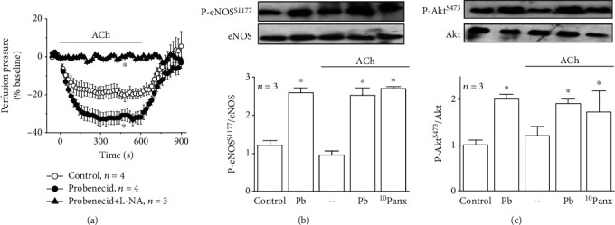
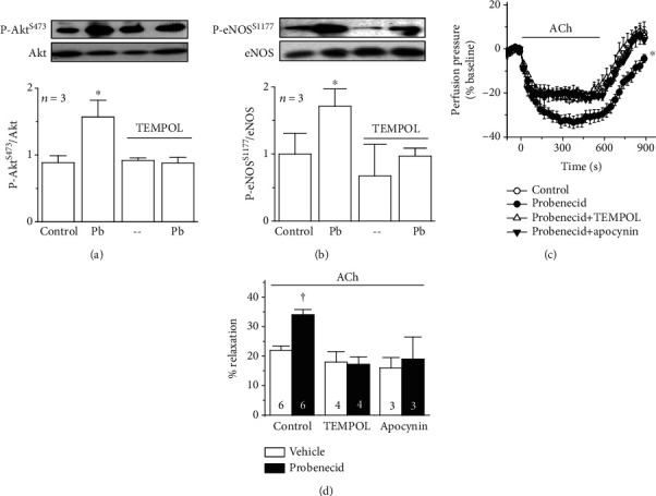
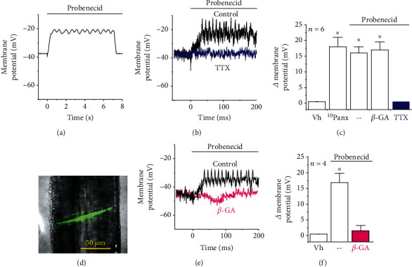
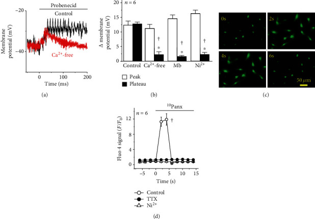
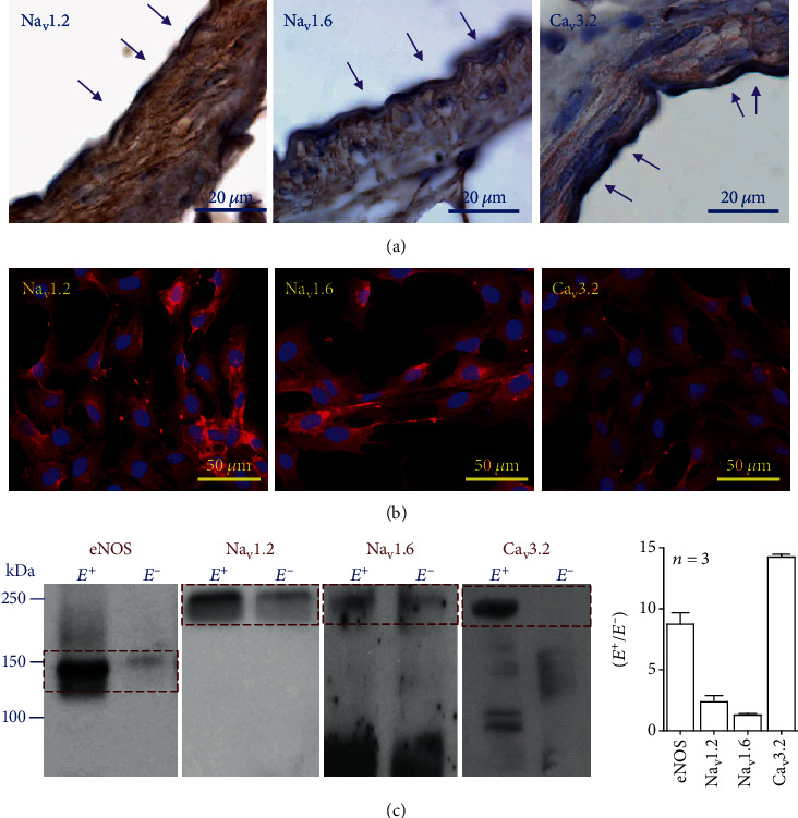
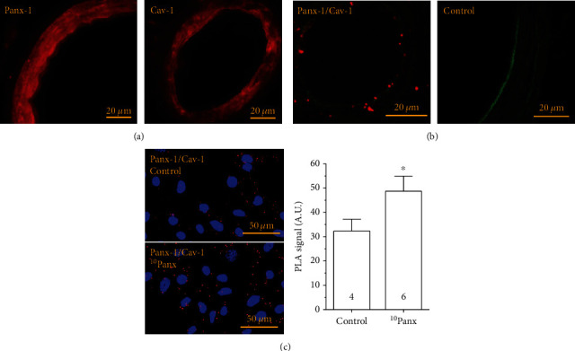
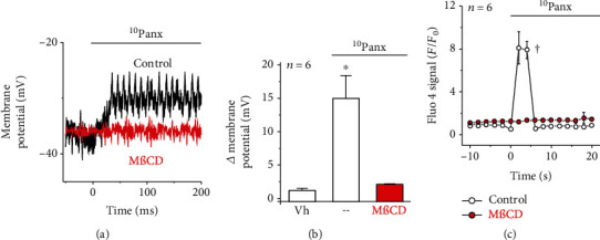
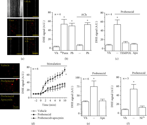
Deletion of pannexin-1 (Panx-1) leads not only to a reduction in endothelium-derived hyperpolarization but also to an increase in NO-mediated vasodilation. Therefore, we evaluated the participation of Panx-1-formed channels in the control of membrane potential and [Ca] of endothelial cells. Changes in NO-mediated vasodilation, membrane potential, superoxide anion (O ) formation, and endothelial cell [Ca] were analyzed in rat isolated mesenteric arterial beds and primary cultures of mesenteric endothelial cells. Inhibition of Panx-1 channels with probenecid (1 mM) or the Panx-1 blocking peptide Panx (60 M) evoked an increase in the ACh (100 nM)-induced vasodilation of KCl-contracted mesenteries and in the phosphorylation level of endothelial NO synthase (eNOS) at serine 1177 (P-eNOS) and Akt at serine 473 (P-Akt). In addition, probenecid or Panx application activated a rapid, tetrodotoxin (TTX, 300 nM)-sensitive, membrane potential depolarization and [Ca] increase in endothelial cells. Interestingly, the endothelial cell depolarization was converted into a transient spike after removing Ca ions from the buffer solution and in the presence of 100 M mibefradil or 10 M Ni. As expected, Ni also abolished the increment in [Ca]. Expression of Na1.2, Na1.6, and Ca3.2 isoforms of voltage-dependent Na and Ca channels was confirmed by immunocytochemistry. Furthermore, the Panx-1 channel blockade was associated with an increase in O production. Treatment with 10 M TEMPOL or 100 M apocynin prevented the increase in O formation, ACh-induced vasodilation, P-eNOS, and P-Akt observed in response to Panx-1 inhibition. These findings indicate that the Panx-1 channel blockade triggers a novel complex signaling pathway initiated by the sequential activation of TTX-sensitive Na channels and Ca3.2 channels, leading to an increase in NO-mediated vasodilation through a NADPH oxidase-dependent P-eNOS, which suggests that Panx-1 may be involved in the endothelium-dependent control of arterial blood pressure.
敲除泛连接蛋白-1(Panx-1)不仅会导致内皮源性超极化减弱,还会使一氧化氮(NO)介导的血管舒张增强。因此,我们评估了由Panx-1形成的通道在内皮细胞膜电位和[Ca]调控中的作用。在大鼠离体肠系膜动脉床和肠系膜内皮细胞原代培养物中,分析了NO介导的血管舒张、膜电位、超氧阴离子(O )生成以及内皮细胞[Ca]的变化。用丙磺舒(1 mM)或Panx-1阻断肽Panx(60 μM)抑制Panx-1通道,可使乙酰胆碱(100 nM)诱导的氯化钾预收缩肠系膜血管舒张增强,同时内皮型一氧化氮合酶(eNOS)丝氨酸1177位点(P-eNOS)和Akt丝氨酸473位点(P-Akt)的磷酸化水平升高。此外,应用丙磺舒或Panx可激活内皮细胞快速的、对河豚毒素(TTX,300 nM)敏感的膜电位去极化和[Ca]升高。有趣的是,在从缓冲溶液中去除钙离子后,以及在存在100 μM米贝拉地尔或10 μM镍的情况下,内皮细胞去极化转变为短暂的尖峰。正如预期的那样,镍也消除了[Ca]的升高。通过免疫细胞化学证实了电压依赖性钠通道和钙通道的Na1.2、Na1.6和Ca3.2亚型的表达。此外,Panx-1通道阻断与O 生成增加有关。用10 μM TEMPOL或100 μM阿朴吗啡处理可防止在Panx-1抑制后观察到的O 生成增加、乙酰胆碱诱导的血管舒张、P-eNOS和P-Akt升高。这些发现表明,Panx-1通道阻断触发了一条由TTX敏感的钠通道和Ca3.2通道依次激活引发的新型复杂信号通路,通过依赖烟酰胺腺嘌呤二核苷酸磷酸(NADPH)氧化酶的P-eNOS导致NO介导的血管舒张增加,这表明Panx-1可能参与动脉血压的内皮依赖性调控。