• 文献检索
  • 文档翻译
  • 深度研究
  • 学术资讯
  • Suppr Zotero 插件Zotero 插件
  • 邀请有礼
  • 套餐&价格
  • 历史记录
应用&插件
Suppr Zotero 插件Zotero 插件浏览器插件Mac 客户端Windows 客户端微信小程序
定价
高级版会员购买积分包购买API积分包
服务
文献检索文档翻译深度研究API 文档MCP 服务
关于我们
关于 Suppr公司介绍联系我们用户协议隐私条款
关注我们

Suppr 超能文献

核心技术专利:CN118964589B侵权必究
粤ICP备2023148730 号-1Suppr @ 2026

文献检索

告别复杂PubMed语法,用中文像聊天一样搜索,搜遍4000万医学文献。AI智能推荐,让科研检索更轻松。

立即免费搜索

文件翻译

保留排版,准确专业,支持PDF/Word/PPT等文件格式,支持 12+语言互译。

免费翻译文档

深度研究

AI帮你快速写综述,25分钟生成高质量综述,智能提取关键信息,辅助科研写作。

立即免费体验

具有眼表面鳞状上皮肿瘤样光学相干断层扫描特征的非恶性结膜上皮肿块。

Non-malignant conjunctival epithelial masses with ocular surface squamous neoplasia-like optical coherence tomography features.

机构信息

Department of Ophthalmology, Semmelweis University, Budapest, Hungary.

2nd Department of Pathology, Semmelweis University, Budapest, Hungary.

出版信息

Int Ophthalmol. 2021 May;41(5):1827-1834. doi: 10.1007/s10792-021-01743-y. Epub 2021 Mar 10.

DOI:10.1007/s10792-021-01743-y
PMID:33694094
原文链接:https://pmc.ncbi.nlm.nih.gov/articles/PMC8087600/
Abstract

PURPOSE

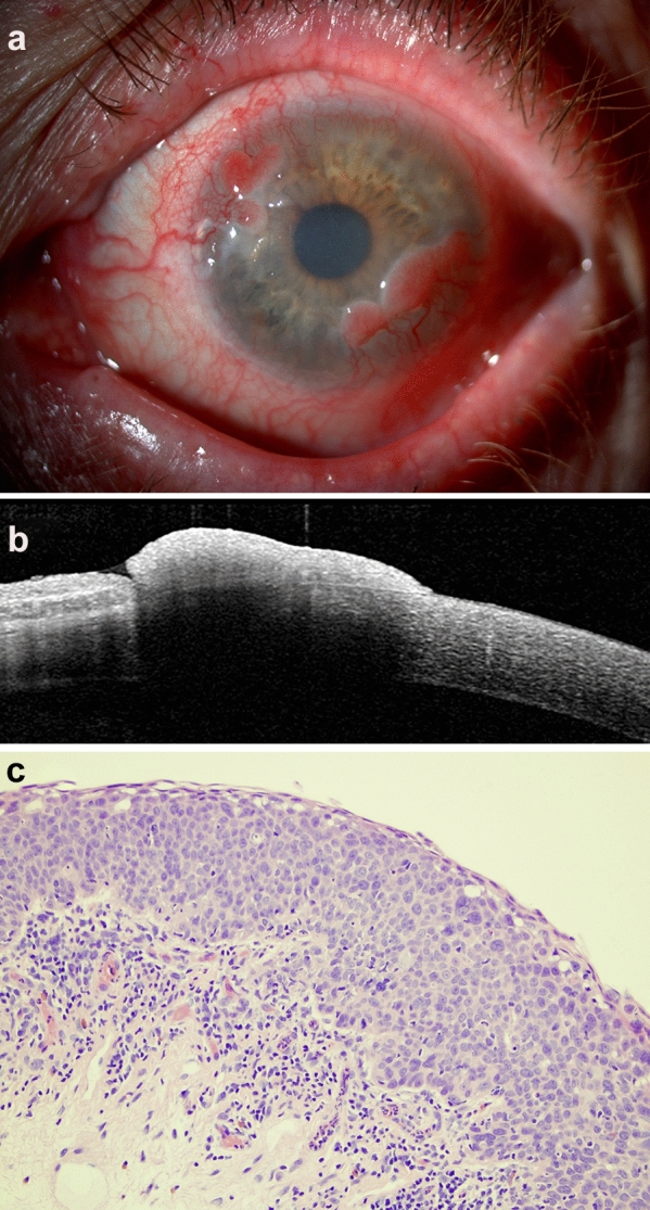
To observe and describe the anterior segment optical coherence tomography features of limbally localised non-malignant epithelial mass lesions METHODS: Thirteen patients (age: 66.9 ± 16.3 years) with conjunctival mass suggesting ocular surface squamous neoplasia with biomicroscopic examination were imaged using anterior segment ocular coherence tomography (anterior segment optical coherence tomography)/Cirrus HD-OCT, Model 4000, Carl Zeiss Meditec, Inc., Dublin, CA, and Spectralis HRA + OCT system, Heidelberg Engineering, Vista, CA/. Cases with ocular surface squamous neoplasia-like anterior segment optical coherence tomography (hyperreflective, thickened epithelium and an abrupt transition from normal to abnormal) were included in the study. Maximal thickness of the epithelium was measured. Histological diagnosis was gained from an excisional or incisional biopsy or impression cytology specimens.

RESULTS

In six patients (age: 68.5 ± 15.4 years) with ocular surface squamous neoplasia-like anterior segment optical coherence tomography features, the histological diagnosis was other than ocular surface squamous neoplasia (papilloma, parakeratosis and a keratotic plaque with mild dysplasia), and ocular surface squamous neoplasia in seven cases (age: 65.6 ± 18.0 years). The maximal epithelial thickness was between 250 and 859 µm in non-ocular surface squamous neoplasia cases and between 252 and 596 µm in ocular surface squamous neoplasia cases.

CONCLUSION

Non-malignant epithelial lesions can mimic ocular surface squamous neoplasia on anterior segment optical coherence tomography.

摘要

目的

观察和描述局限性眼表上皮肿块病变的眼前节光学相干断层扫描特征。

方法

对 13 名(年龄:66.9 ± 16.3 岁)因结膜肿块而怀疑为眼表鳞状上皮肿瘤的患者进行眼前节光学相干断层扫描(前节光学相干断层扫描)/Cirrus HD-OCT、Model 4000、Carl Zeiss Meditec,Inc.,Dublin,CA 和 Spectralis HRA + OCT 系统,Heidelberg Engineering,Vista,CA/成像。本研究纳入了具有眼表鳞状上皮肿瘤样眼前节光学相干断层扫描(高反射性、增厚的上皮和从正常到异常的突然转变)特征的病例。测量上皮的最大厚度。组织学诊断来自切除或切开活检或印模细胞学标本。

结果

在 6 名(年龄:68.5 ± 15.4 岁)具有眼表鳞状上皮肿瘤样眼前节光学相干断层扫描特征的患者中,组织学诊断为非眼表鳞状上皮肿瘤(乳头状瘤、角化不全和角化斑块伴轻度发育不良),而在 7 名患者(年龄:65.6 ± 18.0 岁)中诊断为眼表鳞状上皮肿瘤。非眼表鳞状上皮肿瘤病例的上皮最大厚度在 250 至 859 µm 之间,眼表鳞状上皮肿瘤病例的上皮最大厚度在 252 至 596 µm 之间。

结论

非恶性上皮病变在眼前节光学相干断层扫描上可模拟眼表鳞状上皮肿瘤。

https://cdn.ncbi.nlm.nih.gov/pmc/blobs/3713/8087600/d3881bdad380/10792_2021_1743_Fig2_HTML.jpg
https://cdn.ncbi.nlm.nih.gov/pmc/blobs/3713/8087600/b0457c4b0e80/10792_2021_1743_Fig1_HTML.jpg
https://cdn.ncbi.nlm.nih.gov/pmc/blobs/3713/8087600/d3881bdad380/10792_2021_1743_Fig2_HTML.jpg
https://cdn.ncbi.nlm.nih.gov/pmc/blobs/3713/8087600/b0457c4b0e80/10792_2021_1743_Fig1_HTML.jpg
https://cdn.ncbi.nlm.nih.gov/pmc/blobs/3713/8087600/d3881bdad380/10792_2021_1743_Fig2_HTML.jpg

相似文献

1
Non-malignant conjunctival epithelial masses with ocular surface squamous neoplasia-like optical coherence tomography features.具有眼表面鳞状上皮肿瘤样光学相干断层扫描特征的非恶性结膜上皮肿块。
Int Ophthalmol. 2021 May;41(5):1827-1834. doi: 10.1007/s10792-021-01743-y. Epub 2021 Mar 10.
2
Ultra high-resolution anterior segment optical coherence tomography in the diagnosis and management of ocular surface squamous neoplasia.超高分辨率眼前节光学相干断层扫描在眼表鳞状上皮肿瘤的诊断和治疗中的应用。
Ocul Surf. 2014 Jan;12(1):46-58. doi: 10.1016/j.jtos.2013.11.001. Epub 2013 Nov 9.
3
Anterior segment optical coherence tomography characteristics of conjunctival papilloma as compared to papilliform ocular surface squamous neoplasia.眼前节光学相干断层扫描特征比较结膜乳头瘤与乳头瘤样眼表鳞状上皮瘤。
Eye (Lond). 2023 Apr;37(5):995-1001. doi: 10.1038/s41433-022-02309-7. Epub 2022 Nov 19.
4
Case Series: High-resolution Optical Coherence Tomography as an Optical Biopsy in Ocular Surface Squamous Neoplasia.病例系列:高分辨率光学相干断层扫描在眼表面鳞状上皮肿瘤中的光学活检作用。
Optom Vis Sci. 2021 May 1;98(5):450-455. doi: 10.1097/OPX.0000000000001684.
5
Ultra-high resolution optical coherence tomography for differentiation of ocular surface squamous neoplasia and pterygia.用于区分眼表鳞状上皮肿瘤和翼状胬肉的超高分辨率光学相干断层扫描。
Ophthalmology. 2012 Mar;119(3):481-6. doi: 10.1016/j.ophtha.2011.08.028. Epub 2011 Dec 9.
6
Anterior segment optical coherence tomography in ocular surface tumours and simulating lesions.眼前节光学相干断层扫描在眼表肿瘤和模拟病变中的应用。
Eye (Lond). 2023 Apr;37(5):925-937. doi: 10.1038/s41433-022-02339-1. Epub 2022 Dec 13.
7
High resolution anterior segment optical coherence tomography for differential diagnosis between corneo-conjunctival intraepithelial neoplasia and pterygium.用于角膜结膜上皮内瘤变与翼状胬肉鉴别诊断的高分辨率眼前节光学相干断层扫描
Arch Soc Esp Oftalmol (Engl Ed). 2020 Mar;95(3):108-113. doi: 10.1016/j.oftal.2020.01.002. Epub 2020 Jan 30.
8
Anterior segment optical coherence tomography features of ocular surface squamous epithelial hyperplasia: A report of four cases.眼表鳞状上皮增生的眼前节光学相干断层扫描特征:4例报告
Oman J Ophthalmol. 2023 Jun 27;16(2):314-317. doi: 10.4103/ojo.ojo_173_22. eCollection 2023 May-Aug.
9
How Can We Diagnose Ocular Surface Squamous Neoplasia With Optical Coherence Tomography?我们如何通过光学相干断层扫描诊断眼表鳞状上皮肿瘤?
Cureus. 2023 Mar 17;15(3):e36320. doi: 10.7759/cureus.36320. eCollection 2023 Mar.
10
Anterior segment optical coherence tomography features of pseudoepitheliomatous hyperplasia of the ocular surface: a study of 9 lesions.眼前节光学相干断层扫描在眼表面假上皮瘤样增生中的特征:9 个病变的研究。
Int Ophthalmol. 2021 Jan;41(1):113-119. doi: 10.1007/s10792-020-01558-3. Epub 2020 Aug 25.

引用本文的文献

1
Anterior segment optical coherence tomography in ocular surface tumours and simulating lesions.眼前节光学相干断层扫描在眼表肿瘤和模拟病变中的应用。
Eye (Lond). 2023 Apr;37(5):925-937. doi: 10.1038/s41433-022-02339-1. Epub 2022 Dec 13.
2
Detection of Human Papillomavirus in Squamous Lesions of the Conjunctiva Using RNA and DNA In-Situ Hybridization.应用 RNA 和 DNA 原位杂交技术检测结膜鳞状病变中人乳头瘤病毒。
Int J Mol Sci. 2022 Jun 29;23(13):7249. doi: 10.3390/ijms23137249.

本文引用的文献

1
Update on Diagnosis and Management of Conjunctival Papilloma.结膜乳头状瘤的诊断与管理最新进展
Eye Vis (Lond). 2019 Jun 18;6:18. doi: 10.1186/s40662-019-0142-5. eCollection 2019.
2
High-Resolution Anterior Segment Optical Coherence Tomography in Intraepithelial Versus Invasive Ocular Surface Squamous Neoplasia.高分辨率眼前节光学相干断层扫描在眼表上皮内与浸润性鳞状细胞癌中的应用
Cornea. 2018 Oct;37(10):1292-1298. doi: 10.1097/ICO.0000000000001680.
3
Anterior segment optical coherence tomography.眼前节光学相干断层扫描。
Prog Retin Eye Res. 2018 Sep;66:132-156. doi: 10.1016/j.preteyeres.2018.04.002. Epub 2018 Apr 7.
4
Role of high resolution optical coherence tomography in diagnosing ocular surface squamous neoplasia with coexisting ocular surface diseases.高分辨率光学相干断层扫描在合并眼表疾病的眼表鳞状上皮肿瘤诊断中的作用。
Ocul Surf. 2017 Oct;15(4):688-695. doi: 10.1016/j.jtos.2017.03.003. Epub 2017 Mar 24.
5
Diagnosis and Medical Management of Ocular Surface Squamous Neoplasia.眼表鳞状上皮肿瘤的诊断与药物治疗
Expert Rev Ophthalmol. 2017;12(1):11-19. doi: 10.1080/17469899.2017.1263567. Epub 2016 Dec 5.
6
High-Resolution Optical Coherence Tomography as an Adjunctive Tool in the Diagnosis of Corneal and Conjunctival Pathology.高分辨率光学相干断层扫描作为角膜和结膜病变诊断的辅助工具。
Ocul Surf. 2015 Jul;13(3):226-35. doi: 10.1016/j.jtos.2015.02.001. Epub 2015 Apr 6.
7
Ultra high-resolution anterior segment optical coherence tomography in the diagnosis and management of ocular surface squamous neoplasia.超高分辨率眼前节光学相干断层扫描在眼表鳞状上皮肿瘤的诊断和治疗中的应用。
Ocul Surf. 2014 Jan;12(1):46-58. doi: 10.1016/j.jtos.2013.11.001. Epub 2013 Nov 9.
8
Ultra-high resolution optical coherence tomography for differentiation of ocular surface squamous neoplasia and pterygia.用于区分眼表鳞状上皮肿瘤和翼状胬肉的超高分辨率光学相干断层扫描。
Ophthalmology. 2012 Mar;119(3):481-6. doi: 10.1016/j.ophtha.2011.08.028. Epub 2011 Dec 9.
9
Diagnosis and management of conjunctival and corneal intraepithelial neoplasia using ultra high-resolution optical coherence tomography.使用超高速分辨率光学相干断层扫描技术诊断和治疗结膜和角膜上皮内瘤变。
Ophthalmology. 2011 Aug;118(8):1531-7. doi: 10.1016/j.ophtha.2011.01.005. Epub 2011 Apr 20.
10
Ultra high-resolution anterior segment optical coherence tomography in the evaluation of anterior corneal dystrophies and degenerations.超高分辨率眼前节光学相干断层扫描在评价前部角膜营养不良和变性中的应用。
Ophthalmology. 2011 Jul;118(7):1291-6. doi: 10.1016/j.ophtha.2010.12.015. Epub 2011 Mar 21.