Leibniz-Forschungsinstitut für Molekulare Pharmakologie im Forschungsverbund Berlin e.V. (FMP), Berlin, Germany.
Mildred-Scheel Early Career Center, Medical Faculty, Technische Universität Dresden, Dresden, Germany.
F1000Res. 2020 Nov 26;9:1373. doi: 10.12688/f1000research.27140.2. eCollection 2020.
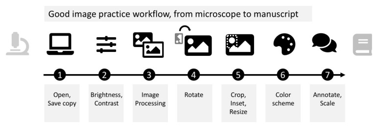
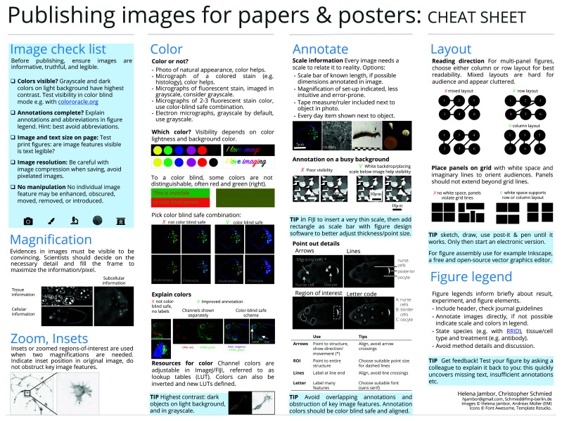
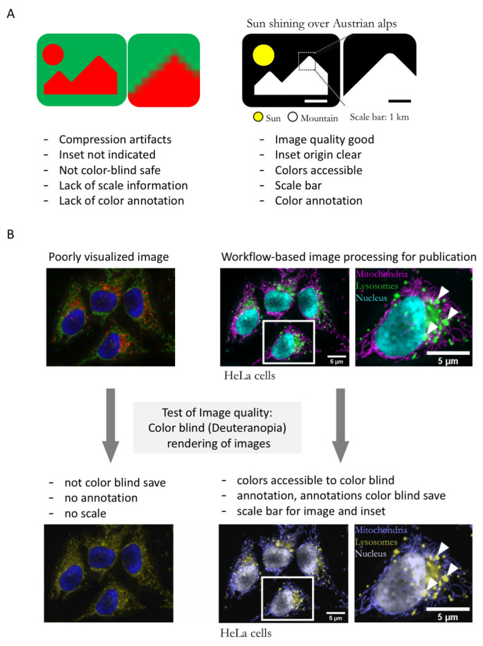
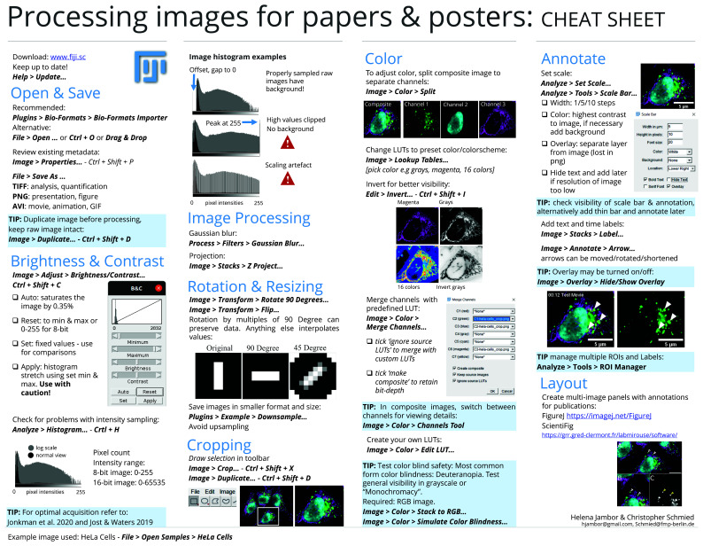
Today, 25% of figures in biomedical publications contain images of various types, e.g. photos, light or electron microscopy images, x-rays, or even sketches or drawings. Despite being widely used, published images may be ineffective or illegible since details are not visible, information is missing or they have been inappropriately processed. The vast majority of such imperfect images can be attributed to the lack of experience of the authors as undergraduate and graduate curricula lack courses on image acquisition, ethical processing, and visualization. Here we present a step-by-step image processing workflow for effective and ethical image presentation. The workflow is aimed to allow novice users with little or no prior experience in image processing to implement the essential steps towards publishing images. The workflow is based on the open source software Fiji, but its principles can be applied with other software packages. All image processing steps discussed here, and complementary suggestions for image presentation, are shown in an accessible "cheat sheet"-style format, enabling wide distribution, use, and adoption to more specific needs.
如今,生物医学出版物中有 25%的内容包含各种类型的图像,例如照片、光学或电子显微镜图像、X 射线,甚至是草图或绘画。尽管被广泛使用,但已发表的图像可能效果不佳或难以辨认,因为细节不可见、信息缺失或处理不当。大多数此类不完美的图像都可以归因于作者缺乏经验,因为本科和研究生课程缺乏关于图像采集、道德处理和可视化的课程。在这里,我们展示了一个用于有效和道德的图像呈现的分步图像处理工作流程。该工作流程旨在允许没有图像处理经验或经验很少的新手用户实施发布图像的基本步骤。该工作流程基于开源软件 Fiji,但它的原理可以应用于其他软件包。这里讨论的所有图像处理步骤,以及有关图像呈现的补充建议,都以易于访问的“备忘单”风格格式呈现,从而能够更广泛地分发、使用和采用更具体的需求。