Department of Ophthalmology, Kitasato University School of Medicine, Sagamihara, Kanagawa, Japan.
Department of Ophthalmology, International University of Health and Welfare Atami Hospital, Atami, Shizuoka, Japan.
PLoS One. 2021 Mar 12;16(3):e0248497. doi: 10.1371/journal.pone.0248497. eCollection 2021.
Age-related distance esotropia (ARDE) involves acquired esotropia at distance and phoria at near. However, distance-independent esotropia (DIE) exists esotropia both at distance and near. Thus, we examined the orbital magnetic resonance imaging (MRI) findings for DIE to assess differences in its characteristics.
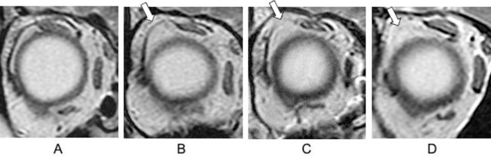
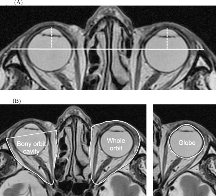
This study was a retrospective case-control study. We evaluated the efficacy of the standard coronal MRI in patients with acquired esotropia and control patients with optic neuritis. Cases with strabismus in the control group were excluded. DIE was defined as having esotropia both at distance and near, and an angle of more than 10 prism diopters at near. The condition of the lateral rectus-superior rectus band, position of rectus muscles, and the volume ratio of the globe to the whole orbit (G/WO) were examined.
The DIE group consisted of 12 eyes of 6 patients (77.3±7.7 years); ARDE group, 38 eyes of 19 patients (73.1±6.8 years); and control group, 34 eyes of 17 patients (70.9±4.3 years). The ratio of abnormality of the lateral rectus-superior rectus bands was higher in the DIE and ARDE groups than in the control group (p<0.01). The vertical angle of the lateral rectus deviated downwards in the control (-7.5±5.1°), ARDE (-12.2±9.1°), and DIE groups (-18.8±5.7°) (p<0.05). The tilting angle of the lateral rectus was tilted temporally in the control (-12.2±9.1°), ARDE (-20.0±8.6°) and DIE groups (-28.6±5.4°) (p<0.01). G/WO was higher in the DIE (0.28±0.01) and ARDE groups (0.27±0.02) compared to the control (0.25±0.03) group (p<0.01).
In comparison with the ARDE and control groups, the DIE group presented with abnormalities of the lateral rectus-superior rectus band, malposition of the lateral rectus, and differences in the G/WO. The DIE group showed a more severe form of ARDE.
年龄相关性外斜视(ARDE)涉及远距获得性外斜视和近距隐斜视。然而,存在与距离无关的外斜视(DIE),即远距和近距均存在外斜视。因此,我们研究了 DIE 的眼眶磁共振成像(MRI)发现,以评估其特征差异。
这是一项回顾性病例对照研究。我们评估了获得性外斜视患者和对照组视神经炎患者标准冠状 MRI 的疗效。对照组中伴有斜视的病例被排除在外。DIE 的定义为远距和近距均有外斜视,近距斜视角度大于 10 棱镜度。检查了外直肌-上直肌带的状况、直肌位置以及眼球与整个眼眶的容积比(G/WO)。
DIE 组由 6 例患者的 12 只眼(77.3±7.7 岁)组成;ARDE 组由 19 例患者的 38 只眼(73.1±6.8 岁)组成;对照组由 17 例患者的 34 只眼(70.9±4.3 岁)组成。DIE 和 ARDE 组的外侧直肌-上直肌带异常比例高于对照组(p<0.01)。对照组(-7.5±5.1°)、ARDE 组(-12.2±9.1°)和 DIE 组(-18.8±5.7°)的外侧直肌垂直角度向下偏斜(p<0.05)。对照组(-12.2±9.1°)、ARDE 组(-20.0±8.6°)和 DIE 组(-28.6±5.4°)的外侧直肌倾斜角度向颞侧倾斜(p<0.01)。DIE(0.28±0.01)和 ARDE(0.27±0.02)组的 G/WO 高于对照组(0.25±0.03)(p<0.01)。
与 ARDE 组和对照组相比,DIE 组外侧直肌-上直肌带异常、外侧直肌位置异常和 G/WO 差异明显。DIE 组表现出更严重的 ARDE 形式。