Department of Radiology, The Second Affiliated Hospital of Harbin Medical University, Harbin, China.
Department of Ultrasound, Harbin the First Hospital, Harbin, China.
Abdom Radiol (NY). 2021 Jul;46(7):3227-3237. doi: 10.1007/s00261-021-03015-w. Epub 2021 Mar 13.
Metastasis-associated in colon cancer 1 (MACC1) and Spondin2 (SPON2) are newly discovered oncogenes, but little is known about their role in colorectal cancer(CRC) liver metastases. PET has become an important molecular imaging technology due to its high sensitivity and quantifiability. In particular, its targeted, specific molecular probes can detect biological behaviors. This study was designed to evaluate the different biological properties of F-FDG, F-FLT, and F-FMISO PET. The value of the CRC liver metastasis model explores the correlation and potential mechanisms of three tracers uptakes with tumor-related biological characteristics.
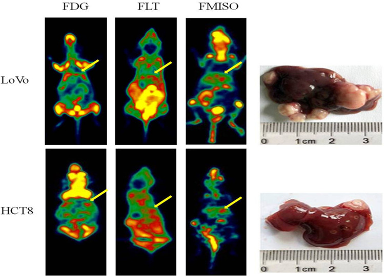
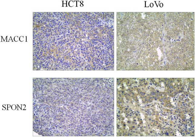
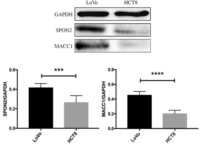
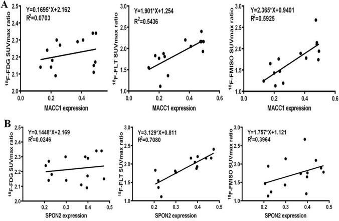
Human CRC cell lines(LoVo and HCT8), were cultured for in vitro radionuclide uptake experiments to compare the molecular imaging features of colorectal cancer cells with different metastatic potentials. Two kinds of cells were injected into the spleen of nude mice to establish a liver metastasis model. After the tumor formation, three kinds of tracer PET images were performed to evaluate the characteristics of live PET imaging of high and low liver metastasis colorectal cancer models. The expression levels of MACC1 and SPON2 in tissues were detected by immunohistochemistry and Western blot. Correlation between tracer uptake and expression of MACC1 and SPON2 in liver metastases was assessed by linear regression analysis.
The uptake rate of in vitro three tracers uptake experiments was LoVo > HCT8. Micro-PET scan showed no significant difference between the F-FDG SUV values of the two cells (P > 0.05); there was significant difference between the F-FLT and F-FMISO SUV values (P < 0.05). All in vivo FLT and FMISO SUV values were significantly higher in LoVo tumors than in HCT8 tumors. The results of Western blot and immunohistochemistry showed that the expression levels of MACC1 and SPON2 in LoVo liver metastasis were higher than those in HCT8 (P < 0.05). The F-FLT SUVmax ratio was significantly correlated with the expression of MACC1 and SPON2 in hepatic metastases (r = 0.737, P = 0.0026; r = 0.842, P = 0.0002). The F-FMISO SUVmax ratio was only significantly correlated with the expression of MACC1 in hepatic metastasis (r = 0.770, P = 0.0013).
Early screening with F-FLT and F-FMISO tracers has important clinical value for the efficient diagnosis and treatment of colorectal cancer liver metastases.
结肠癌转移相关基因 1(MACC1)和 Spondin2(SPON2)是新发现的癌基因,但它们在结直肠癌肝转移中的作用知之甚少。正电子发射断层扫描(PET)已成为一种重要的分子成像技术,因为它具有高灵敏度和可量化性。特别是,其靶向、特异性分子探针可以检测生物行为。本研究旨在评估 F-FDG、F-FLT 和 F-FMISO PET 的不同生物学特性。利用结直肠癌肝转移模型探讨三种示踪剂摄取与肿瘤相关生物学特征的相关性及潜在机制。
体外放射性核素摄取实验培养人结直肠癌细胞系(LoVo 和 HCT8),比较不同转移潜能的结直肠癌细胞的分子成像特征。将两种细胞注入裸鼠脾脏建立肝转移模型。肿瘤形成后,行三种示踪剂 PET 图像评价高低转移潜能结直肠癌肝转移模型的活体 PET 成像特征。免疫组化和 Western blot 检测组织中 MACC1 和 SPON2 的表达水平。线性回归分析评估示踪剂摄取与肝转移中 MACC1 和 SPON2 表达的相关性。
体外三种示踪剂摄取实验的摄取率为 LoVo>HCT8。微 PET 扫描显示两种细胞的 F-FDG SUV 值无显著差异(P>0.05);F-FLT 和 F-FMISO SUV 值有显著差异(P<0.05)。LoVo 肿瘤的所有体内 FLT 和 FMISO SUV 值均显著高于 HCT8 肿瘤。Western blot 和免疫组化结果显示,LoVo 肝转移组织中 MACC1 和 SPON2 的表达水平高于 HCT8(P<0.05)。F-FLT SUVmax 比值与肝转移组织中 MACC1 和 SPON2 的表达呈显著正相关(r=0.737,P=0.0026;r=0.842,P=0.0002)。F-FMISO SUVmax 比值仅与肝转移组织中 MACC1 的表达呈显著正相关(r=0.770,P=0.0013)。
F-FLT 和 F-FMISO 示踪剂的早期筛查对结直肠癌肝转移的有效诊断和治疗具有重要的临床价值。