Niemann Annika, Voß Samuel, Tulamo Riikka, Weigand Simon, Preim Bernhard, Berg Philipp, Saalfeld Sylvia
Faculty of Computer Science, Otto-von-Guericke University Magdeburg, Universitätsplatz 2, D-39106, Magdeburg, Germany.
Laboratory of Fluid Dynamics and Technical Flows, Otto-von-Guericke University Magdeburg, Magdeburg, Germany.
Int J Comput Assist Radiol Surg. 2021 Apr;16(4):597-607. doi: 10.1007/s11548-021-02334-z. Epub 2021 Mar 14.
For the evaluation and rupture risk assessment of intracranial aneurysms, clinical, morphological and hemodynamic parameters are analyzed. The reliability of intracranial hemodynamic simulations strongly depends on the underlying models. Due to the missing information about the intracranial vessel wall, the patient-specific wall thickness is often neglected as well as the specific physiological and pathological properties of the vessel wall.
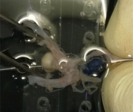
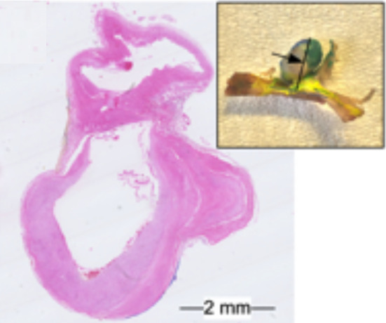
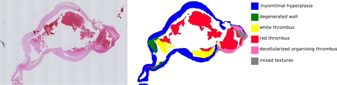
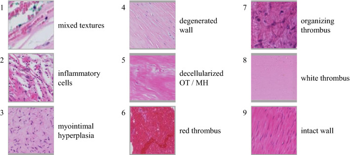
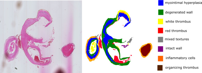
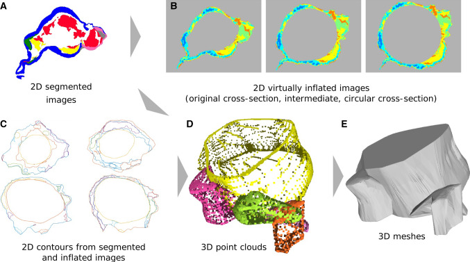
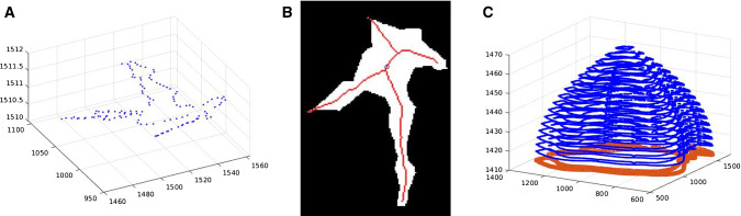
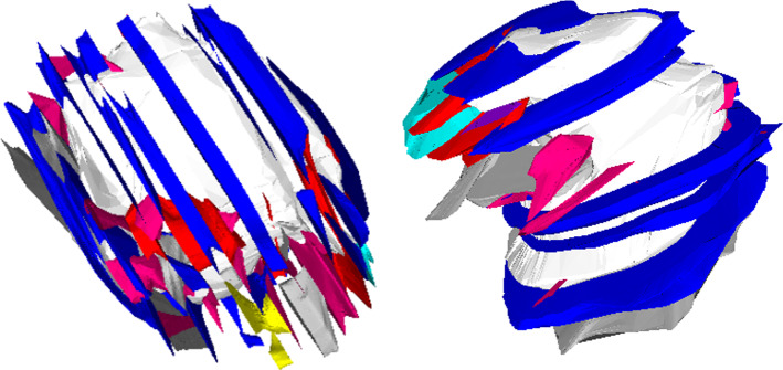
In this work, we present a model for structural simulations with patient-specific wall thickness including different tissue types based on postmortem histologic image data. Images of histologic 2D slices from intracranial aneurysms were manually segmented in nine tissue classes. After virtual inflation, they were combined into 3D models. This approach yields multiple 3D models of the inner and outer wall and different tissue parts as a prerequisite for subsequent simulations.
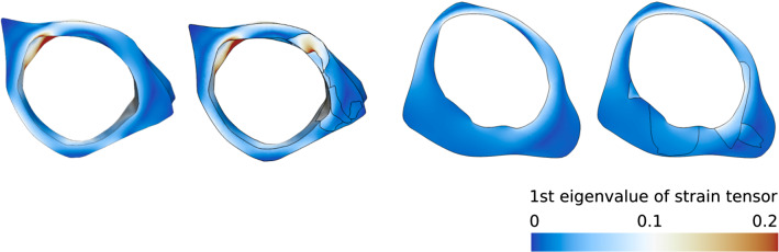
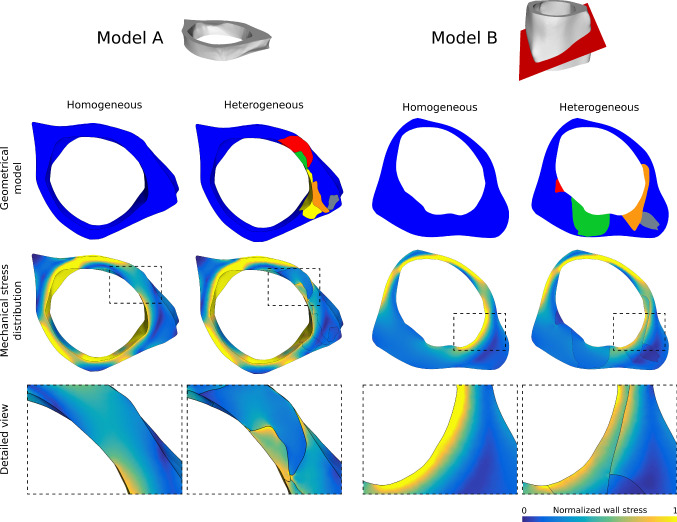
We presented a pipeline to generate 3D models of aneurysms with respect to the different tissue textures occurring in the wall. First experiments show that including the variance of the tissue in the structural simulation affect the simulation result. Especially at the interfaces between neighboring tissue classes, the larger influence of stiffer components on the stability equilibrium became obvious.
The presented approach enables the creation of a geometric model with differentiated wall tissue. This information can be used for different applications, like hemodynamic simulations, to increase the modeling accuracy.
为评估颅内动脉瘤及破裂风险,对临床、形态学和血流动力学参数进行分析。颅内血流动力学模拟的可靠性很大程度上取决于基础模型。由于缺乏关于颅内血管壁的信息,患者特异性的壁厚以及血管壁的特定生理和病理特性常常被忽视。
在这项工作中,我们基于死后组织学图像数据,提出了一种针对具有患者特异性壁厚且包含不同组织类型的结构模拟模型。来自颅内动脉瘤的组织学二维切片图像被手动分割为九种组织类别。经过虚拟充气后,它们被组合成三维模型。这种方法产生了多个内壁、外壁和不同组织部分的三维模型,作为后续模拟的前提条件。
我们提出了一个管道,用于生成针对血管壁中出现的不同组织纹理的动脉瘤三维模型。初步实验表明,在结构模拟中纳入组织的差异会影响模拟结果。特别是在相邻组织类别之间的界面处,较硬成分对稳定性平衡的更大影响变得明显。
所提出的方法能够创建具有差异化壁组织的几何模型。这些信息可用于不同的应用,如血流动力学模拟,以提高建模精度。