Department of Microbiology, Immunology and Physiology, School of Medicine, Meharry Medical College, Nashville, TN, United States.
School of Graduate Studies and Research, Meharry Medical College, Nashville, TN, United States.
Front Immunol. 2021 Feb 25;12:607044. doi: 10.3389/fimmu.2021.607044. eCollection 2021.
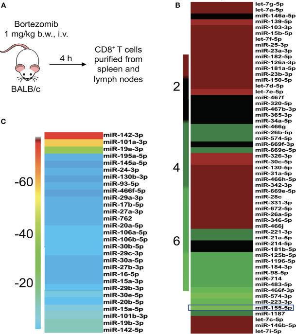
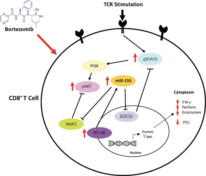
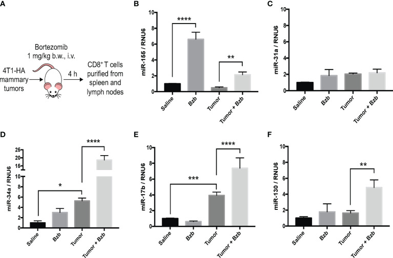
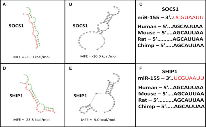
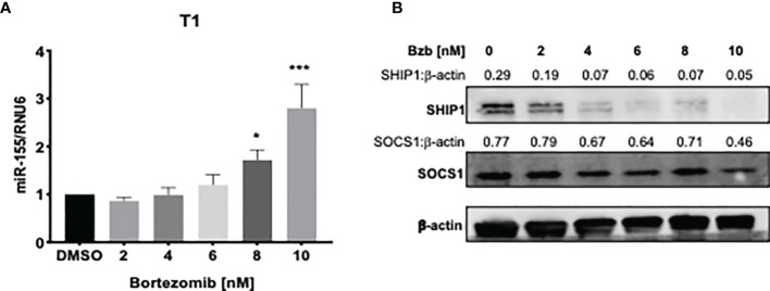
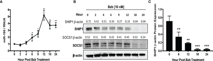
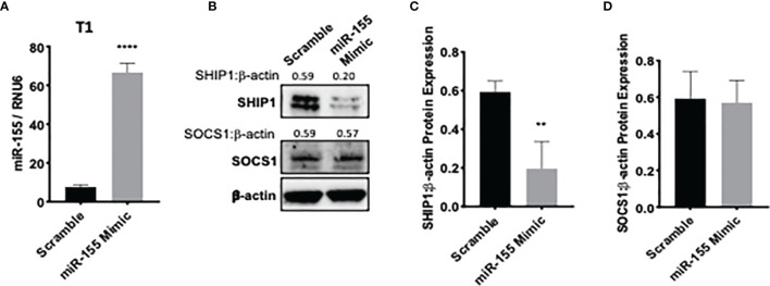
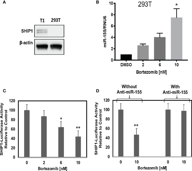
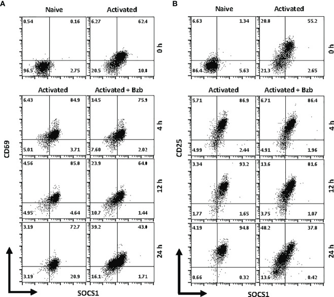
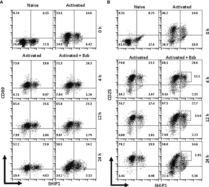
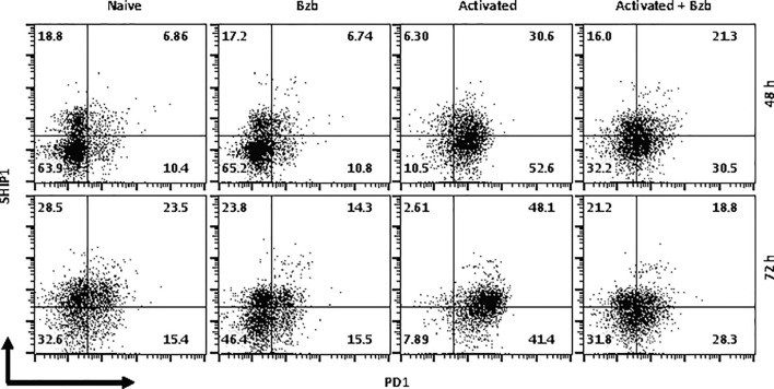
Suppressive mechanisms operating within T cells are linked to immune dysfunction in the tumor microenvironment. We have previously reported using adoptive T cell immunotherapy models that tumor-bearing mice treated with a regimen of proteasome inhibitor, bortezomib - a dipeptidyl boronate, show increased antitumor lymphocyte effector function and survival. Here, we identify a mechanism for the improved antitumor CD8 T cell function following bortezomib treatment. Intravenous administration of bortezomib at a low dose (1 mg/kg body weight) in wild-type or tumor-bearing mice altered the expression of a number of miRNAs in CD8 T cells. Specifically, the effect of bortezomib was prominent on miR-155 - a key cellular miRNA involved in T cell function. Importantly, bortezomib-induced upregulation of miR-155 was associated with the downregulation of its targets, the suppressor of cytokine signaling 1 (SOCS1) and inositol polyphosphate-5-phosphatase (SHIP1). Genetic and biochemical analysis confirmed a functional link between miR-155 and these targets. Moreover, activated CD8 T cells treated with bortezomib exhibited a significant reduction in programmed cell death-1 (PD-1) expressing SHIP1 phenotype. These data underscore a mechanism of action by which bortezomib induces miR-155-dependent downregulation of SOCS1 and SHIP1 negative regulatory proteins, leading to a suppressed PD-1-mediated T cell exhaustion. Collectively, data provide novel molecular insights into bortezomib-mediated lymphocyte-stimulatory effects that could overcome immunosuppressive actions of tumor on antitumor T cell functions. The findings support the approach that bortezomib combined with other immunotherapies would lead to improved therapeutic outcomes by overcoming T cell exhaustion in the tumor microenvironment.
抑制性机制在 T 细胞中发挥作用与肿瘤微环境中的免疫功能障碍有关。我们之前曾报道过,在使用过继性 T 细胞免疫治疗模型时,接受蛋白酶体抑制剂硼替佐米治疗的荷瘤小鼠表现出增强的抗肿瘤淋巴细胞效应功能和存活。在这里,我们确定了硼替佐米治疗后抗肿瘤 CD8 T 细胞功能改善的机制。在野生型或荷瘤小鼠中静脉给予低剂量(1mg/kg 体重)的硼替佐米会改变 CD8 T 细胞中许多 miRNA 的表达。具体而言,硼替佐米对 miR-155 的作用尤为明显 - miR-155 是一种参与 T 细胞功能的关键细胞 miRNA。重要的是,硼替佐米诱导的 miR-155 上调与靶基因的下调有关,即细胞因子信号转导抑制因子 1(SOCS1)和肌醇多磷酸-5-磷酸酶(SHIP1)。遗传和生化分析证实了 miR-155 与这些靶基因之间的功能联系。此外,用硼替佐米处理的激活的 CD8 T 细胞表现出程序性细胞死亡-1(PD-1)表达的 SHIP1 表型显著减少。这些数据强调了硼替佐米诱导 miR-155 依赖性下调 SOCS1 和 SHIP1 负调节蛋白的作用机制,导致 PD-1 介导的 T 细胞衰竭受到抑制。总之,数据为硼替佐米介导的淋巴细胞刺激作用提供了新的分子见解,这种作用可能克服肿瘤对抗肿瘤 T 细胞功能的免疫抑制作用。研究结果支持硼替佐米与其他免疫疗法联合使用的方法,通过克服肿瘤微环境中的 T 细胞衰竭,可改善治疗效果。