Wei Qiaolin, Arami Hamed, Santos Hélder A, Zhang Hongbo, Li Yangyang, He Jian, Zhong Danni, Ling Daishun, Zhou Min
The Fourth Affiliated Hospital Zhejiang University School of Medicine Yiwu 322000 P. R. China.
Institute of Translational Medicine Zhejiang University Hangzhou 310009 P. R. China.
Adv Sci (Weinh). 2021 Jan 18;8(5):2002788. doi: 10.1002/advs.202002788. eCollection 2021 Mar.
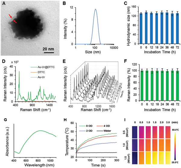
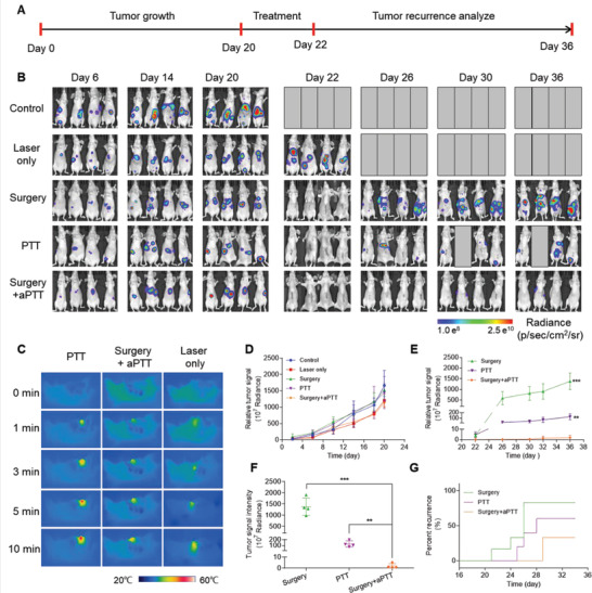
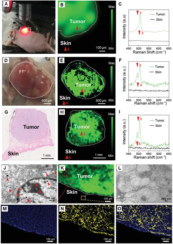
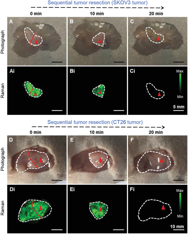
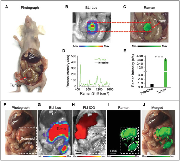
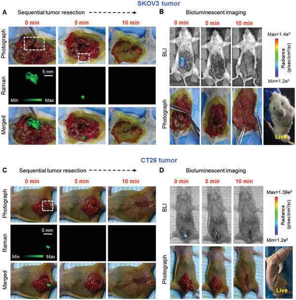
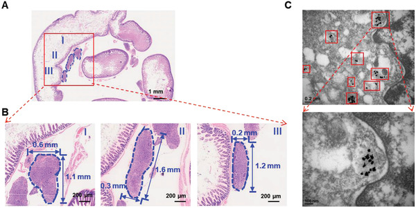
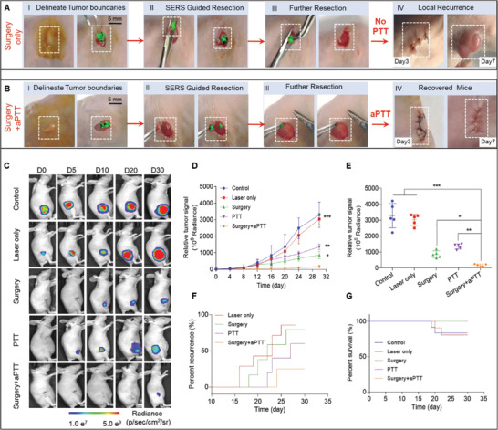
Surgical resection is commonly used for therapeutic management of different solid tumors and is regarded as a primary standard of care procedure, but precise localization of tumor margins is a major intraoperative challenge. Herein, a generalized method by optimizing gold nanoparticles for intraoperative detection and photothermal ablation of tumor margins is introduced. These nanoparticles are detectable by highly sensitive surface-enhanced Raman scattering imaging. This non-invasive technique assists in delineating the two surgically challenged tumors in live mice with orthotopic colon or ovarian tumors. Any remaining residual tumors are also ablated by using post-surgical adjuvant photothermaltherapy (aPTT), which results in microscale heat generation due to interaction of these nanoparticles with near-infrared laser. Ablation of these post-operative residual micro-tumors prolongs the survival of mice significantly and delays tumor recurrence by 15 days. To validate clinical translatability of this method, the pharmacokinetics, biodistribution, Raman contrast, aPTT efficiency, and toxicity of these nanoparticles are also investigated. The nanoparticles have long blood circulation time (≈24 h), high tumor accumulation (4.87 ± 1.73%ID g) and no toxicity. This high-resolution and sensitive intraoperative approach is versatile and can be potentially used for targeted ablation of residual tumor after resection within different organs.
手术切除常用于不同实体瘤的治疗管理,被视为主要的标准治疗程序,但肿瘤边缘的精确定位是术中的一项重大挑战。在此,介绍一种通过优化金纳米颗粒用于术中检测和肿瘤边缘光热消融的通用方法。这些纳米颗粒可通过高灵敏度表面增强拉曼散射成像进行检测。这种非侵入性技术有助于在患有原位结肠或卵巢肿瘤的活体小鼠中勾勒出两个手术难度较大的肿瘤。任何残留的肿瘤也可通过术后辅助光热疗法(aPTT)进行消融,由于这些纳米颗粒与近红外激光相互作用,会产生微尺度的热量。消融这些术后残留的微肿瘤可显著延长小鼠的生存期,并将肿瘤复发延迟15天。为验证该方法的临床可转化性,还对这些纳米颗粒的药代动力学、生物分布、拉曼对比度、aPTT效率和毒性进行了研究。这些纳米颗粒具有较长的血液循环时间(约24小时)、高肿瘤蓄积(4.87±1.73%ID/g)且无毒性。这种高分辨率且灵敏的术中方法具有通用性,可潜在地用于不同器官切除术后残留肿瘤的靶向消融。