• 文献检索
  • 文档翻译
  • 深度研究
  • 学术资讯
  • Suppr Zotero 插件Zotero 插件
  • 邀请有礼
  • 套餐&价格
  • 历史记录
应用&插件
Suppr Zotero 插件Zotero 插件浏览器插件Mac 客户端Windows 客户端微信小程序
定价
高级版会员购买积分包购买API积分包
服务
文献检索文档翻译深度研究API 文档MCP 服务
关于我们
关于 Suppr公司介绍联系我们用户协议隐私条款
关注我们

Suppr 超能文献

核心技术专利:CN118964589B侵权必究
粤ICP备2023148730 号-1Suppr @ 2026

文献检索

告别复杂PubMed语法,用中文像聊天一样搜索,搜遍4000万医学文献。AI智能推荐,让科研检索更轻松。

立即免费搜索

文件翻译

保留排版,准确专业,支持PDF/Word/PPT等文件格式,支持 12+语言互译。

免费翻译文档

深度研究

AI帮你快速写综述,25分钟生成高质量综述,智能提取关键信息,辅助科研写作。

立即免费体验

β-分泌酶1(BACE1)对损伤的周围神经中巨噬细胞募集和活性的影响。

The influence of BACE1 on macrophage recruitment and activity in the injured peripheral nerve.

作者信息

Fissel John A, Farah Mohamed H

机构信息

Department of Neurology, Johns Hopkins University School of Medicine, The John G. Rangos Sr. Building, Room 239, 855 N. Wolfe Street, Baltimore, MD, 21205, USA.

出版信息

J Neuroinflammation. 2021 Mar 15;18(1):71. doi: 10.1186/s12974-021-02121-2.

DOI:10.1186/s12974-021-02121-2
PMID:33722254
原文链接:https://pmc.ncbi.nlm.nih.gov/articles/PMC7962400/
Abstract

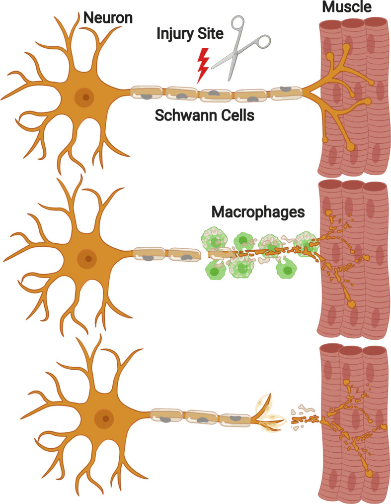
Following peripheral nerve injury, multiple cell types, including axons, Schwann cells, and macrophages, coordinate to promote nerve regeneration. However, this capacity for repair is limited, particularly in older populations, and current treatments are insufficient. A critical component of the regeneration response is the network of cell-to-cell signaling in the injured nerve microenvironment. Sheddases are expressed in the peripheral nerve and play a role in the regulation if this cell-to-cell signaling through cleavage of transmembrane proteins, enabling the regulation of multiple pathways through cis- and trans-cellular regulatory mechanisms. Enhanced axonal regeneration has been observed in mice with deletion of the sheddase beta-secretase (BACE1), a transmembrane aspartyl protease that has been studied in the context of Alzheimer's disease. BACE1 knockout (KO) mice display enhanced macrophage recruitment and activity following nerve injury, although it is unclear whether this plays a role in driving the enhanced axonal regeneration. Further, it is unknown by what mechanism(s) BACE1 increases macrophage recruitment and activity. BACE1 has many substrates, several of which are known to have immunomodulatory activity. This review will discuss current knowledge of the role of BACE1 and other sheddases in peripheral nerve regeneration and outline known immunomodulatory BACE1 substrates and what potential roles they could play in peripheral nerve regeneration. Currently, the literature suggests that BACE1 and substrates that are expressed by neurons and Schwann cells are likely to be more important for this process than those expressed by macrophages. More broadly, BACE1 may play a role as an effector of immunomodulation beyond the peripheral nerve.

摘要

外周神经损伤后,多种细胞类型,包括轴突、施万细胞和巨噬细胞,协同促进神经再生。然而,这种修复能力是有限的,尤其是在老年人群中,而且目前的治疗方法并不充分。再生反应的一个关键组成部分是受损神经微环境中的细胞间信号网络。去整合素金属蛋白酶在周围神经中表达,并通过切割跨膜蛋白在调节这种细胞间信号中发挥作用,从而能够通过顺式和反式细胞调节机制调节多种途径。在缺失去整合素金属蛋白酶β-分泌酶(BACE1)的小鼠中观察到轴突再生增强,BACE1是一种跨膜天冬氨酸蛋白酶,已在阿尔茨海默病的背景下进行了研究。BACE1基因敲除(KO)小鼠在神经损伤后显示出巨噬细胞募集和活性增强,尽管尚不清楚这是否在驱动轴突再生增强中发挥作用。此外,BACE1通过何种机制增加巨噬细胞募集和活性尚不清楚。BACE1有许多底物,其中一些已知具有免疫调节活性。本综述将讨论目前关于BACE1和其他去整合素金属蛋白酶在周围神经再生中的作用的知识,并概述已知的免疫调节性BACE1底物以及它们在周围神经再生中可能发挥的潜在作用。目前,文献表明,神经元和施万细胞表达的BACE1和底物可能比巨噬细胞表达的底物在这个过程中更重要。更广泛地说,BACE1可能在周围神经之外作为免疫调节的效应器发挥作用。

https://cdn.ncbi.nlm.nih.gov/pmc/blobs/9bfc/7962400/9af83c44345e/12974_2021_2121_Fig4_HTML.jpg
https://cdn.ncbi.nlm.nih.gov/pmc/blobs/9bfc/7962400/f639b5516704/12974_2021_2121_Fig1_HTML.jpg
https://cdn.ncbi.nlm.nih.gov/pmc/blobs/9bfc/7962400/a53c11670c82/12974_2021_2121_Fig2_HTML.jpg
https://cdn.ncbi.nlm.nih.gov/pmc/blobs/9bfc/7962400/7d18ee120f3d/12974_2021_2121_Fig3_HTML.jpg
https://cdn.ncbi.nlm.nih.gov/pmc/blobs/9bfc/7962400/9af83c44345e/12974_2021_2121_Fig4_HTML.jpg

相似文献

1
The influence of BACE1 on macrophage recruitment and activity in the injured peripheral nerve.β-分泌酶1(BACE1)对损伤的周围神经中巨噬细胞募集和活性的影响。
J Neuroinflammation. 2021 Mar 15;18(1):71. doi: 10.1186/s12974-021-02121-2.
2
Macrophage-specific deletion of BACE1 does not enhance macrophage recruitment to the injured peripheral nerve.BACE1 特异性缺失并不增强巨噬细胞向损伤周围神经的募集。
J Neuroimmunol. 2020 Dec 15;349:577423. doi: 10.1016/j.jneuroim.2020.577423. Epub 2020 Oct 6.
3
Axonal and Schwann cell BACE1 is equally required for remyelination of peripheral nerves.轴突和施万细胞中的β-分泌酶1(BACE1)对于周围神经的髓鞘再生同样不可或缺。
J Neurosci. 2015 Mar 4;35(9):3806-14. doi: 10.1523/JNEUROSCI.5207-14.2015.
4
Reduced BACE1 activity enhances clearance of myelin debris and regeneration of axons in the injured peripheral nervous system.BACE1 活性降低可增强损伤外周神经系统中髓鞘碎片的清除和轴突的再生。
J Neurosci. 2011 Apr 13;31(15):5744-54. doi: 10.1523/JNEUROSCI.6810-10.2011.
5
Increased BACE1 activity inhibits peripheral nerve regeneration after injury.BACE1 活性增加会抑制损伤后的周围神经再生。
Neurobiol Dis. 2017 Oct;106:147-157. doi: 10.1016/j.nbd.2017.07.003. Epub 2017 Jul 5.
6
Increased TNFR1 expression and signaling in injured peripheral nerves of mice with reduced BACE1 activity.BACE1 活性降低的小鼠损伤外周神经中 TNFR1 表达和信号的增加。
Neurobiol Dis. 2016 Sep;93:21-7. doi: 10.1016/j.nbd.2016.04.002. Epub 2016 Apr 11.
7
Pharmacological BACE Inhibition Improves Axonal Regeneration in Nerve Injury and Disease Models.药物抑制 BACE 可改善神经损伤和疾病模型中的轴突再生。
Neurotherapeutics. 2020 Jul;17(3):973-988. doi: 10.1007/s13311-020-00852-3.
8
Genetic deletion of BACE1 in mice affects remyelination of sciatic nerves.小鼠中β-分泌酶1(BACE1)的基因缺失影响坐骨神经的髓鞘再生。
FASEB J. 2008 Aug;22(8):2970-80. doi: 10.1096/fj.08-106666. Epub 2008 Apr 15.
9
BACE1 influences debris clearance and axonal regeneration in injured peripheral nerve.BACE1 影响损伤外周神经中的碎片清除和轴突再生。
J Peripher Nerv Syst. 2012 Dec;17 Suppl 3:30-3. doi: 10.1111/j.1529-8027.2012.00428.x.
10
Role of macrophages in Wallerian degeneration and axonal regeneration after peripheral nerve injury.巨噬细胞在外周神经损伤后的 Wallerian 变性和轴突再生中的作用。
Acta Neuropathol. 2015 Nov;130(5):605-18. doi: 10.1007/s00401-015-1482-4. Epub 2015 Sep 29.

引用本文的文献

1
The Dance Between Schwann Cells and Macrophages During the Repair of Peripheral Nerve Injury.周围神经损伤修复过程中施万细胞与巨噬细胞之间的相互作用
Neurosci Bull. 2025 Jun 7. doi: 10.1007/s12264-025-01427-y.
2
Molecular and cellular mechanisms underlying peripheral nerve injury-induced cellular ecological shifts: Implications for neuroregeneration.外周神经损伤诱导细胞生态转变的分子和细胞机制:对神经再生的启示
IBRO Neurosci Rep. 2024 Dec 28;18:120-129. doi: 10.1016/j.ibneur.2024.12.013. eCollection 2025 Jun.
3
Biomimetic ECM nerve guidance conduit with dynamic 3D interconnected porous network and sustained IGF-1 delivery for enhanced peripheral nerve regeneration and immune modulation.

本文引用的文献

1
Macrophage-specific deletion of BACE1 does not enhance macrophage recruitment to the injured peripheral nerve.BACE1 特异性缺失并不增强巨噬细胞向损伤周围神经的募集。
J Neuroimmunol. 2020 Dec 15;349:577423. doi: 10.1016/j.jneuroim.2020.577423. Epub 2020 Oct 6.
2
Pharmacological BACE Inhibition Improves Axonal Regeneration in Nerve Injury and Disease Models.药物抑制 BACE 可改善神经损伤和疾病模型中的轴突再生。
Neurotherapeutics. 2020 Jul;17(3):973-988. doi: 10.1007/s13311-020-00852-3.
3
Dominant mutations of the Notch ligand Jagged1 cause peripheral neuropathy.
具有动态三维互连多孔网络和持续递送胰岛素样生长因子-1的仿生细胞外基质神经导管,用于增强周围神经再生和免疫调节。
Mater Today Bio. 2024 Dec 12;30:101403. doi: 10.1016/j.mtbio.2024.101403. eCollection 2025 Feb.
4
A Review and Bibliometric Analysis of Studies on Advances in Peripheral Nerve Regeneration.外周神经再生进展研究的综述与文献计量分析
Cureus. 2024 Sep 16;16(9):e69515. doi: 10.7759/cureus.69515. eCollection 2024 Sep.
5
Single-cell and spatial multiomic inference of gene regulatory networks using SCRIPro.使用SCRIPro进行基因调控网络的单细胞和空间多组学推断
Bioinformatics. 2024 Jul 18;40(7). doi: 10.1093/bioinformatics/btae466.
6
Enhanced sciatic nerve regeneration by relieving iron-overloading and organelle stress with the nanofibrous P(MMD-co-LA)/DFO conduits.通过纳米纤维P(MMD-co-LA)/DFO导管减轻铁过载和细胞器应激来增强坐骨神经再生。
Mater Today Bio. 2022 Aug 6;16:100387. doi: 10.1016/j.mtbio.2022.100387. eCollection 2022 Dec.
7
Peripheral Nerve Injury Treatments and Advances: One Health Perspective.周围神经损伤的治疗与进展:从“One Health”角度来看。
Int J Mol Sci. 2022 Jan 14;23(2):918. doi: 10.3390/ijms23020918.
8
Application of Hybrid Electrically Conductive Hydrogels Promotes Peripheral Nerve Regeneration.混合导电水凝胶的应用促进周围神经再生。
Gels. 2022 Jan 6;8(1):41. doi: 10.3390/gels8010041.
Notch 配体 Jagged1 的显性突变导致周围神经病。
J Clin Invest. 2020 Mar 2;130(3):1506-1512. doi: 10.1172/JCI128152.
4
Deletion of SIRPα (signal regulatory protein-α) promotes phagocytic clearance of myelin debris in Wallerian degeneration, axon regeneration, and recovery from nerve injury.SIRPα(信号调节蛋白-α)缺失可促进 Wallerian 变性、轴突再生和神经损伤恢复过程中髓鞘碎片的吞噬清除。
J Neuroinflammation. 2019 Dec 28;16(1):277. doi: 10.1186/s12974-019-1679-x.
5
Interleukin-1 and Related Cytokines in the Regulation of Inflammation and Immunity.白细胞介素-1 及其相关细胞因子在炎症和免疫调节中的作用。
Immunity. 2019 Apr 16;50(4):778-795. doi: 10.1016/j.immuni.2019.03.012.
6
The Complex Work of Proteases and Secretases in Wallerian Degeneration: Beyond Neuregulin-1.沃勒变性中蛋白酶和分泌酶的复杂作用:超越神经调节蛋白-1
Front Cell Neurosci. 2019 Mar 20;13:93. doi: 10.3389/fncel.2019.00093. eCollection 2019.
7
The Success and Failure of the Schwann Cell Response to Nerve Injury.施万细胞对神经损伤反应的成功与失败
Front Cell Neurosci. 2019 Feb 11;13:33. doi: 10.3389/fncel.2019.00033. eCollection 2019.
8
Mixed Lineage Kinase Domain-like Protein MLKL Breaks Down Myelin following Nerve Injury.混合谱系激酶结构域样蛋白 MLKL 破坏神经损伤后的髓鞘。
Mol Cell. 2018 Nov 1;72(3):457-468.e5. doi: 10.1016/j.molcel.2018.09.011. Epub 2018 Oct 18.
9
Experiments on the Section of the Glosso-Pharyngeal and Hypoglossal Nerves of the Frog, and Observations of the Alterations Produced Thereby in the Structure of Their Primitive Fibres.关于青蛙舌咽神经和舌下神经切断的实验,以及对由此引起的其原始纤维结构变化的观察
Edinb Med Surg J. 1851 Oct 1;76(189):369-376.
10
Macrophages Regulate Schwann Cell Maturation after Nerve Injury.巨噬细胞调控神经损伤后的雪旺细胞成熟。
Cell Rep. 2018 Sep 4;24(10):2561-2572.e6. doi: 10.1016/j.celrep.2018.08.004.