Bauer Nicholas C, Yang Anli, Wang Xin, Zhou Yunli, Klibanski Anne, Soberman Roy J
Division of Nephrology, Department of Medicine, Massachusetts General Hospital and Harvard Medical School, Charlestown, Massachusetts, United States.
Neuroendocrine Unit, Department of Medicine, Massachusetts General Hospital and Harvard Medical School, Boston, Massachusetts, United States.
J Biol Chem. 2021 Jan-Jun;296:100540. doi: 10.1016/j.jbc.2021.100540. Epub 2021 Mar 12.
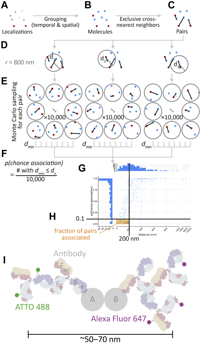
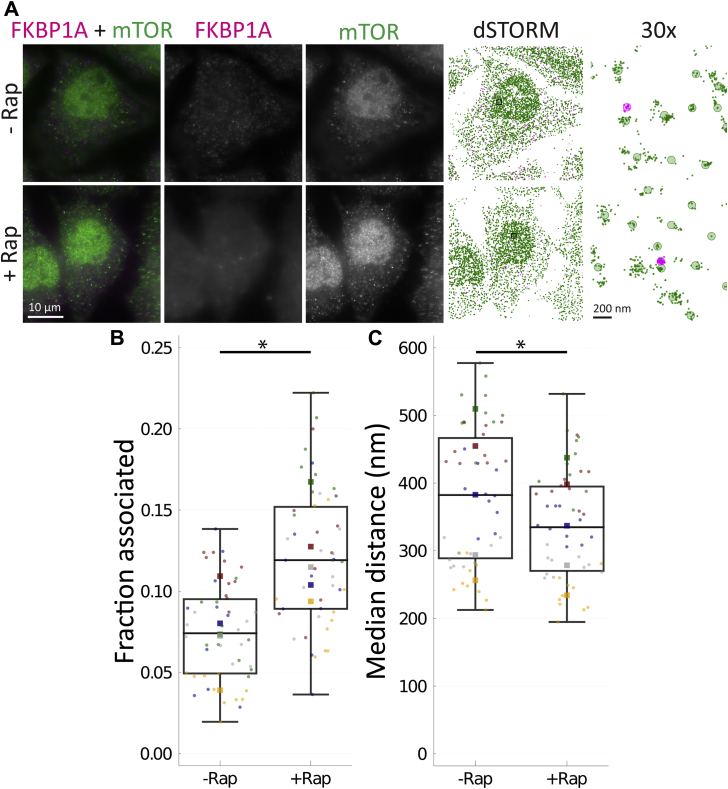
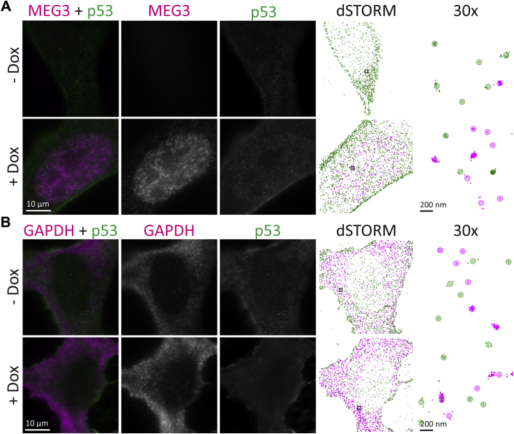
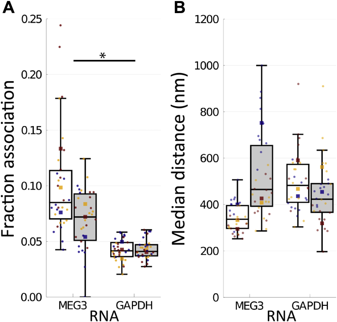
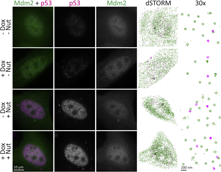
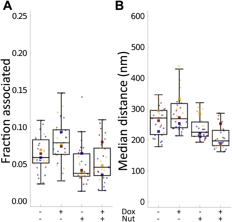
The functions of long noncoding (lnc)RNAs, such as MEG3, are defined by their interactions with other RNAs and proteins. These interactions, in turn, are shaped by their subcellular localization and temporal context. Therefore, it is important to be able to analyze the relationships of lncRNAs while preserving cellular architecture. The ability of MEG3 to suppress cell proliferation led to its recognition as a tumor suppressor. MEG3 has been proposed to activate p53 by disrupting the interaction of p53 with mouse double minute 2 homolog (Mdm2). To test this mechanism in the native cellular context, we employed two-color direct stochastic optical reconstruction microscopy, a single-molecule localization microscopy technique, to detect and quantify the localizations of p53, Mdm2, and MEG3 in U2OS cells. We developed a new cross-nearest neighbor/Monte Carlo algorithm to quantify the association of these molecules. Proof of concept for our method was obtained by examining the association between FKBP1A and mTOR, MEG3 and p53, and Mdm2 and p53. In contrast to previous models, our data support a model in which MEG3 modulates p53 independently of the interaction with Mdm2.
长链非编码(lnc)RNA(如MEG3)的功能是由它们与其他RNA和蛋白质的相互作用所决定的。反过来,这些相互作用又受到它们的亚细胞定位和时间背景的影响。因此,在保留细胞结构的同时能够分析lncRNA之间的关系非常重要。MEG3抑制细胞增殖的能力使其被视为一种肿瘤抑制因子。有人提出MEG3通过破坏p53与小鼠双微体2同源物(Mdm2)的相互作用来激活p53。为了在天然细胞环境中验证这一机制,我们采用了双色直接随机光学重建显微镜(一种单分子定位显微镜技术)来检测和量化U2OS细胞中p53、Mdm2和MEG3的定位。我们开发了一种新的交叉最近邻/蒙特卡罗算法来量化这些分子之间的关联。通过检测FKBP1A与mTOR、MEG3与p53以及Mdm2与p53之间的关联,我们获得了该方法的概念验证。与之前的模型不同,我们的数据支持一种MEG3独立于与Mdm2的相互作用来调节p53的模型。