• 文献检索
  • 文档翻译
  • 深度研究
  • 学术资讯
  • Suppr Zotero 插件Zotero 插件
  • 邀请有礼
  • 套餐&价格
  • 历史记录
应用&插件
Suppr Zotero 插件Zotero 插件浏览器插件Mac 客户端Windows 客户端微信小程序
定价
高级版会员购买积分包购买API积分包
服务
文献检索文档翻译深度研究API 文档MCP 服务
关于我们
关于 Suppr公司介绍联系我们用户协议隐私条款
关注我们

Suppr 超能文献

核心技术专利:CN118964589B侵权必究
粤ICP备2023148730 号-1Suppr @ 2026

文献检索

告别复杂PubMed语法,用中文像聊天一样搜索,搜遍4000万医学文献。AI智能推荐,让科研检索更轻松。

立即免费搜索

文件翻译

保留排版,准确专业,支持PDF/Word/PPT等文件格式,支持 12+语言互译。

免费翻译文档

深度研究

AI帮你快速写综述,25分钟生成高质量综述,智能提取关键信息,辅助科研写作。

立即免费体验

定量分析对比增强光谱乳腺摄影灰度值对乳腺癌新辅助化疗病理反应的早期预测。

Quantitative analysis of contrast enhanced spectral mammography grey value for early prediction of pathological response of breast cancer to neoadjuvant chemotherapy.

机构信息

Department of Radiology, Yantai Yuhuangding Hospital, Affiliated Hospital of Qingdao University, No. 20 Yuhuangding East Road, Yantai, 264000, Shandong, People's Republic of China.

GE Healthcare, Institute of Precision Medicine, No. 1 Huatuo Road, Shanghai, 201203, People's Republic of China.

出版信息

Sci Rep. 2021 Mar 15;11(1):5892. doi: 10.1038/s41598-021-85353-9.

DOI:10.1038/s41598-021-85353-9
PMID:33723322
原文链接:https://pmc.ncbi.nlm.nih.gov/articles/PMC7960703/
Abstract

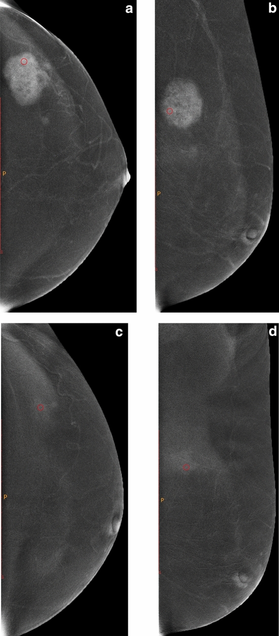
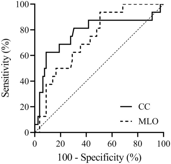
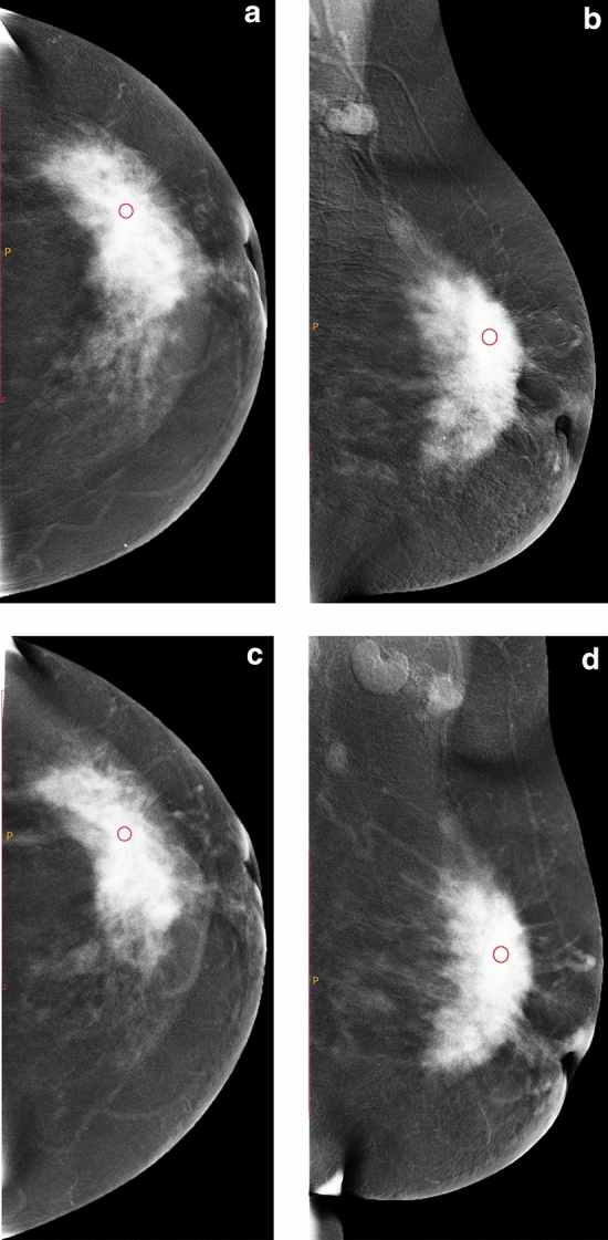
A quantitative analysis of contrast-enhanced spectral mammography (CESM) enhancement was conducted for the early prediction of the pathological response after neoadjuvant chemotherapy (NAC). Retrospective analysis of the data of 111 patients was conducted, and all of them underwent NAC in our hospital and surgical resection after the end of all cycles from January 2018 to May 2019. They were divided into pathological complete response (PCR) and non-PCR groups. We determined whether a statistical difference in the percentage of CESM grey value reduction (ΔCGV) was present in the PCR and non-PCR groups and whether a statistical difference was observed in the diagnostic efficiency of craniocaudal (CC) and mediolateral oblique (MLO) view subtraction images. Independent sample t-test was used to compare different groups, the receiver operating characteristic (ROC) curve was used to compare the diagnostic efficacy of CC and MLO for pathological response after NAC, and the Delong test was used to compare the area under the ROC curve (AUC). Statistical significance was considered at P < 0.05. A statistical difference was observed in the ΔCGV in the PCR and non-PCR groups. No statistical difference was observed in the AUCs of CC and MLO view subtraction images. The ΔCGV can be used as a quantitative index to predict PCR early, and no statistical difference was observed in the diagnostic efficacy of CC and MLO view subtraction images.

摘要

对对比增强光谱乳腺摄影(CESM)增强进行定量分析,以预测新辅助化疗(NAC)后病理反应。回顾性分析了 2018 年 1 月至 2019 年 5 月在我院接受 NAC 并在所有周期结束后进行手术切除的 111 例患者的数据。他们被分为病理完全缓解(PCR)组和非-PCR 组。我们确定了 PCR 组和非-PCR 组之间 CESM 灰度值减少百分比(ΔCGV)是否存在统计学差异,以及头尾位(CC)和内外斜位(MLO)减影图像的诊断效率是否存在统计学差异。采用独立样本 t 检验比较不同组,采用受试者工作特征(ROC)曲线比较 CC 和 MLO 对 NAC 后病理反应的诊断效能,并采用 Delong 检验比较 ROC 曲线下面积(AUC)。P<0.05 为差异有统计学意义。PCR 组和非-PCR 组的ΔCGV 存在统计学差异。CC 和 MLO 位减影图像的 AUC 无统计学差异。ΔCGV 可作为预测 PCR 的定量指标,CC 和 MLO 位减影图像的诊断效能无统计学差异。

https://cdn.ncbi.nlm.nih.gov/pmc/blobs/6973/7960703/2aa8d3740d44/41598_2021_85353_Fig3_HTML.jpg
https://cdn.ncbi.nlm.nih.gov/pmc/blobs/6973/7960703/795b0cc096d4/41598_2021_85353_Fig1_HTML.jpg
https://cdn.ncbi.nlm.nih.gov/pmc/blobs/6973/7960703/f61d4ee5b919/41598_2021_85353_Fig2_HTML.jpg
https://cdn.ncbi.nlm.nih.gov/pmc/blobs/6973/7960703/2aa8d3740d44/41598_2021_85353_Fig3_HTML.jpg
https://cdn.ncbi.nlm.nih.gov/pmc/blobs/6973/7960703/795b0cc096d4/41598_2021_85353_Fig1_HTML.jpg
https://cdn.ncbi.nlm.nih.gov/pmc/blobs/6973/7960703/f61d4ee5b919/41598_2021_85353_Fig2_HTML.jpg
https://cdn.ncbi.nlm.nih.gov/pmc/blobs/6973/7960703/2aa8d3740d44/41598_2021_85353_Fig3_HTML.jpg

相似文献

1
Quantitative analysis of contrast enhanced spectral mammography grey value for early prediction of pathological response of breast cancer to neoadjuvant chemotherapy.定量分析对比增强光谱乳腺摄影灰度值对乳腺癌新辅助化疗病理反应的早期预测。
Sci Rep. 2021 Mar 15;11(1):5892. doi: 10.1038/s41598-021-85353-9.
2
Radiomics of contrast-enhanced spectral mammography for prediction of pathological complete response to neoadjuvant chemotherapy in breast cancer.对比增强光谱乳腺摄影的放射组学分析用于预测乳腺癌新辅助化疗的病理完全缓解。
J Xray Sci Technol. 2023;31(4):669-683. doi: 10.3233/XST-221349.
3
Contrast-enhanced spectral mammography in neoadjuvant chemotherapy monitoring: a comparison with breast magnetic resonance imaging.对比增强光谱乳腺摄影术在新辅助化疗监测中的应用:与乳腺磁共振成像的比较。
Breast Cancer Res. 2017 Sep 11;19(1):106. doi: 10.1186/s13058-017-0899-1.
4
[Texture analysis based on contrast-enhanced MRI can predict treatment response to neoadjuvant chemotherapy of breast cancer].基于对比增强磁共振成像的纹理分析可预测乳腺癌新辅助化疗的治疗反应
Zhonghua Zhong Liu Za Zhi. 2017 May 23;39(5):344-349. doi: 10.3760/cma.j.issn.0253-3766.2017.05.005.
5
Diagnostic Value of Radiomics Analysis in Contrast-Enhanced Spectral Mammography for Identifying Triple-Negative Breast Cancer.基于对比增强光谱乳腺摄影的影像组学分析在三阴性乳腺癌诊断中的价值
Front Oncol. 2021 Dec 23;11:773196. doi: 10.3389/fonc.2021.773196. eCollection 2021.
6
Contrast-Enhanced Spectral Mammography Assessment of Patients Treated with Neoadjuvant Chemotherapy for Breast Cancer.对比增强光谱乳腺摄影评估接受新辅助化疗的乳腺癌患者。
Curr Oncol. 2021 Sep 6;28(5):3448-3462. doi: 10.3390/curroncol28050298.
7
The diagnostic performance of CESM and CE-MRI in evaluating the pathological response to neoadjuvant therapy in breast cancer: a systematic review and meta-analysis.对比增强乳腺X线摄影(CESM)和对比增强磁共振成像(CE-MRI)在评估乳腺癌新辅助治疗病理反应中的诊断性能:一项系统评价和荟萃分析
Br J Radiol. 2020 Aug;93(1112):20200301. doi: 10.1259/bjr.20200301. Epub 2020 Jul 2.
8
Contrast-Enhanced Spectral Mammography Versus Ultrasonography: Diagnostic Performance in Symptomatic Patients with Dense Breasts.对比增强频谱钼靶 X 线摄影与超声检查:致密型乳腺中症状患者的诊断性能。
Korean J Radiol. 2020 Apr;21(4):442-449. doi: 10.3348/kjr.2019.0393.
9
Intratumoral and peritumoral radiomics for preoperative prediction of neoadjuvant chemotherapy effect in breast cancer based on contrast-enhanced spectral mammography.基于对比增强光谱乳腺摄影术的瘤内和瘤周放射组学用于乳腺癌新辅助化疗效果的术前预测
Eur Radiol. 2022 May;32(5):3207-3219. doi: 10.1007/s00330-021-08414-7. Epub 2022 Jan 23.
10
Quantitative contrast-enhanced ultrasound evaluation of pathological complete response in patients with locally advanced breast cancer receiving neoadjuvant chemotherapy.定量对比增强超声评价局部晚期乳腺癌患者接受新辅助化疗后的病理完全缓解。
Eur J Radiol. 2018 Jun;103:118-123. doi: 10.1016/j.ejrad.2018.04.005. Epub 2018 Apr 9.

引用本文的文献

1
Contrast-enhanced Mammography for Early Prediction of Neoadjuvant Therapy Response in Breast Cancer.对比增强乳腺摄影术用于早期预测乳腺癌新辅助治疗反应
Radiol Imaging Cancer. 2025 Jul;7(4):e250244. doi: 10.1148/rycan.250244.
2
Exploring Neoadjuvant Chemotherapy, Predictive Models, Radiomic, and Pathological Markers in Breast Cancer: A Comprehensive Review.探索乳腺癌的新辅助化疗、预测模型、放射组学和病理标志物:一项综述
Cancers (Basel). 2023 Nov 4;15(21):5288. doi: 10.3390/cancers15215288.
3
Neoadjuvant chemotherapy for breast cancer: an evaluation of its efficacy and research progress.

本文引用的文献

1
Radiomic Analysis in Contrast-Enhanced Spectral Mammography for Predicting Breast Cancer Histological Outcome.对比增强光谱乳腺摄影术的放射组学分析用于预测乳腺癌组织学结果
Diagnostics (Basel). 2020 Sep 17;10(9):708. doi: 10.3390/diagnostics10090708.
2
Which clinical, radiological, histological, and molecular parameters are associated with the absence of enhancement of known breast cancers with Contrast Enhanced Digital Mammography (CEDM)?哪些临床、放射学、组织学和分子参数与使用对比增强数字乳腺摄影术(CEDM)的已知乳腺癌无增强有关?
Breast. 2020 Dec;54:15-24. doi: 10.1016/j.breast.2020.08.009. Epub 2020 Aug 21.
3
乳腺癌新辅助化疗:疗效评估及研究进展
Front Oncol. 2023 Oct 3;13:1169010. doi: 10.3389/fonc.2023.1169010. eCollection 2023.
4
A spatial attention guided deep learning system for prediction of pathological complete response using breast cancer histopathology images.基于空间注意力的深度学习系统,用于预测乳腺癌组织病理学图像的病理完全缓解。
Bioinformatics. 2022 Sep 30;38(19):4605-4612. doi: 10.1093/bioinformatics/btac558.
5
Advances in Imaging in Evaluating the Efficacy of Neoadjuvant Chemotherapy for Breast Cancer.评估乳腺癌新辅助化疗疗效的影像学进展
Front Oncol. 2022 May 20;12:816297. doi: 10.3389/fonc.2022.816297. eCollection 2022.
6
Contrast-Enhanced Spectral Mammography Assessment of Patients Treated with Neoadjuvant Chemotherapy for Breast Cancer.对比增强光谱乳腺摄影评估接受新辅助化疗的乳腺癌患者。
Curr Oncol. 2021 Sep 6;28(5):3448-3462. doi: 10.3390/curroncol28050298.
Magnetic resonance imaging in breast cancer management in the context of neo-adjuvant chemotherapy.
磁共振成像在新辅助化疗背景下的乳腺癌管理中的应用。
Crit Rev Oncol Hematol. 2018 Dec;132:51-65. doi: 10.1016/j.critrevonc.2018.09.012. Epub 2018 Sep 25.
4
Quantification of enhancement in contrast-enhanced spectral mammography using a custom-made quantifier tool (I-STRIP): A proof-of-concept study.使用定制定量工具(I-STRIP)对对比增强光谱乳腺摄影中的增强进行定量:概念验证研究。
Eur J Radiol. 2018 Sep;106:114-121. doi: 10.1016/j.ejrad.2018.07.021. Epub 2018 Jul 24.
5
Comparison of Contrast-Enhanced Mammography and Contrast-Enhanced Breast MR Imaging.对比增强乳腺钼靶摄影与对比增强乳腺磁共振成像的比较。
Magn Reson Imaging Clin N Am. 2018 May;26(2):259-263. doi: 10.1016/j.mric.2017.12.005. Epub 2018 Feb 21.
6
Role of MR Imaging in Neoadjuvant Therapy Monitoring.磁共振成像在新辅助治疗监测中的作用。
Magn Reson Imaging Clin N Am. 2018 May;26(2):207-220. doi: 10.1016/j.mric.2017.12.011. Epub 2018 Mar 2.
7
Contrast-Enhanced Spectral Mammography is Comparable to MRI in the Assessment of Residual Breast Cancer Following Neoadjuvant Systemic Therapy.对比增强光谱乳腺成像在新辅助全身治疗后残余乳腺癌评估方面可与 MRI 相媲美。
Ann Surg Oncol. 2018 May;25(5):1350-1356. doi: 10.1245/s10434-018-6413-x. Epub 2018 Mar 7.
8
Contrast-enhanced spectral mammography in neoadjuvant chemotherapy monitoring: a comparison with breast magnetic resonance imaging.对比增强光谱乳腺摄影术在新辅助化疗监测中的应用:与乳腺磁共振成像的比较。
Breast Cancer Res. 2017 Sep 11;19(1):106. doi: 10.1186/s13058-017-0899-1.
9
Contrast-enhanced spectral mammography (CESM) versus breast magnetic resonance imaging (MRI): A retrospective comparison in 66 breast lesions.对比增强光谱乳腺摄影(CESM)与乳腺磁共振成像(MRI):66例乳腺病变的回顾性比较
Diagn Interv Imaging. 2017 Feb;98(2):113-123. doi: 10.1016/j.diii.2016.08.013. Epub 2016 Sep 26.
10
Selective elimination of breast cancer surgery in exceptional responders: historical perspective and current trials.特殊反应者中乳腺癌手术的选择性消除:历史回顾与当前试验
Breast Cancer Res. 2016 Mar 8;18(1):28. doi: 10.1186/s13058-016-0684-6.