Davis Nicola, Mota Bibiana C, Stead Larissa, Palmer Emily O C, Lombardero Laura, Rodríguez-Puertas Rafael, de Paola Vincenzo, Barnes Samuel J, Sastre Magdalena
Department of Brain Sciences, Imperial College London, Hammersmith Hospital, London, W12 0NN, UK.
Department of Pharmacology, University of the Basque Country (UPV/EHU), 48940, Leioa, Spain.
J Neuroinflammation. 2021 Mar 17;18(1):73. doi: 10.1186/s12974-021-02117-y.
Astrocytes provide a vital support to neurons in normal and pathological conditions. In Alzheimer's disease (AD) brains, reactive astrocytes have been found surrounding amyloid plaques, forming an astrocytic scar. However, their role and potential mechanisms whereby they affect neuroinflammation, amyloid pathology, and synaptic density in AD remain unclear.
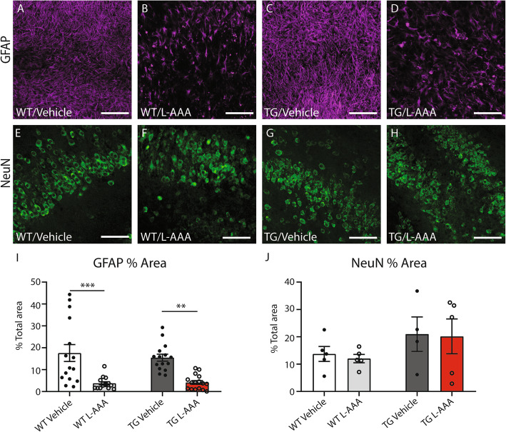
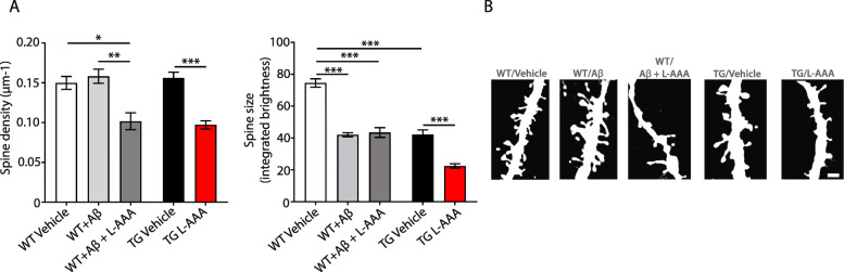
To explore the role of astrocytes on Aβ pathology and neuroinflammatory markers, we pharmacologically ablated them in organotypic brain culture slices (OBCSs) from 5XFAD mouse model of AD and wild-type (WT) littermates with the selective astrocytic toxin L-alpha-aminoadipate (L-AAA). To examine the effects on synaptic circuitry, we measured dendritic spine number and size in OBCSs from Thy-1-GFP transgenic mice incubated with synthetic Aβ42 or double transgenics Thy-1-GFP/5XFAD mice treated with LAAA or vehicle for 24 h.
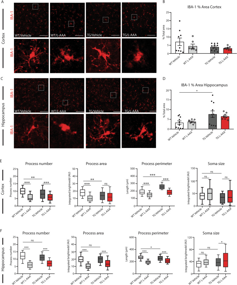
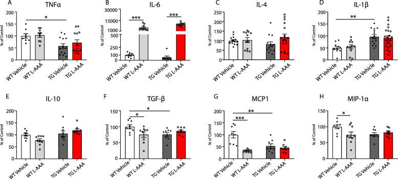
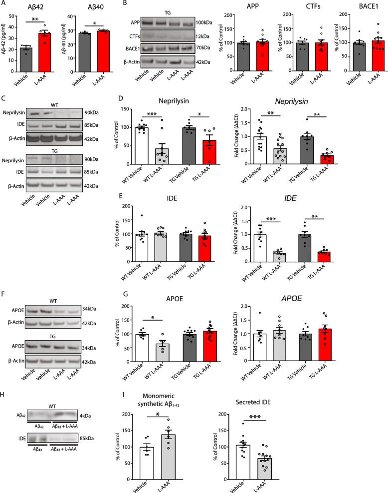
Treatment of OBCSs with L-AAA resulted in an increased expression of pro-inflammatory cytokine IL-6 in conditioned media of WTs and 5XFAD slices, associated with changes in microglia morphology but not in density. The profile of inflammatory markers following astrocytic loss was different in WT and transgenic cultures, showing reductions in inflammatory mediators produced in astrocytes only in WT sections. In addition, pharmacological ablation of astrocytes led to an increase in Aβ levels in homogenates of OBCS from 5XFAD mice compared with vehicle controls, with reduced enzymatic degradation of Aβ due to lower neprilysin and insulin-degrading enzyme (IDE) expression. Furthermore, OBSCs from wild-type mice treated with L-AAA and synthetic amyloid presented 56% higher levels of Aβ in culture media compared to sections treated with Aβ alone, concomitant with reduced expression of IDE in culture medium, suggesting that astrocytes contribute to Aβ clearance and degradation. Quantification of hippocampal dendritic spines revealed a reduction in their density following L-AAA treatment in all groups analyzed. In addition, pharmacological ablation of astrocytes resulted in a decrease in spine size in 5XFAD OBCSs but not in OBCSs from WT treated with synthetic Aβ compared to vehicle control.
Astrocytes play a protective role in AD by aiding Aβ clearance and supporting synaptic plasticity.
在正常和病理条件下,星形胶质细胞为神经元提供至关重要的支持。在阿尔茨海默病(AD)患者的大脑中,已发现反应性星形胶质细胞围绕着淀粉样斑块,形成星形胶质细胞瘢痕。然而,它们在AD中影响神经炎症、淀粉样病理和突触密度的作用及潜在机制仍不清楚。
为了探究星形胶质细胞对Aβ病理和神经炎症标志物的作用,我们用选择性星形胶质细胞毒素L-α-氨基己二酸(L-AAA),在来自AD的5XFAD小鼠模型和野生型(WT)同窝小鼠的脑器官型培养切片(OBCSs)中对星形胶质细胞进行药物性消融。为了检测对突触回路的影响,我们测量了用合成Aβ42孵育的Thy-1-GFP转基因小鼠或用LAAA或溶剂处理24小时的双转基因Thy-1-GFP/5XFAD小鼠的OBCSs中的树突棘数量和大小。
用L-AAA处理OBCSs导致WT和5XFAD切片的条件培养基中促炎细胞因子IL-6的表达增加,这与小胶质细胞形态的改变有关,但与密度无关。星形胶质细胞缺失后炎症标志物的情况在WT和转基因培养物中有所不同,仅在WT切片中显示星形胶质细胞产生的炎症介质减少。此外,与溶剂对照相比,对星形胶质细胞进行药物性消融导致5XFAD小鼠的OBCS匀浆中Aβ水平升高,由于脑啡肽酶和胰岛素降解酶(IDE)表达降低,Aβ的酶促降解减少。此外,与仅用Aβ处理的切片相比,用L-AAA和合成淀粉样蛋白处理的野生型小鼠的OBSCs在培养基中的Aβ水平高56%,同时培养基中IDE的表达降低,这表明星形胶质细胞有助于Aβ的清除和降解。对海马树突棘的定量分析显示,在所有分析的组中,L-AAA处理后其密度降低。此外,与溶剂对照相比,对星形胶质细胞进行药物性消融导致5XFAD OBCSs中的棘突大小减小,但在用合成Aβ处理的WT的OBCSs中没有减小。
星形胶质细胞通过帮助清除Aβ和支持突触可塑性在AD中发挥保护作用。